{"title":"Increased Osteoblast GαS Promotes Ossification by Suppressing Cartilage and Enhancing Callus Mineralization During Fracture Repair in Mice","authors":"Kathy K Lee, Adele Changoor, Marc D Grynpas, Jane Mitchell","doi":"10.1002/jbm4.10841","DOIUrl":null,"url":null,"abstract":"<p>Gα<sub>S</sub>, the stimulatory G protein α-subunit that raises intracellular cAMP levels by activating adenylyl cyclase, plays a vital role in bone development, maintenance, and remodeling. Previously, using transgenic mice overexpressing Gα<sub>S</sub> in osteoblasts (G<sub>S</sub>-Tg), we demonstrated the influence of osteoblast Gα<sub>S</sub> level on osteogenesis, bone turnover, and skeletal responses to hyperparathyroidism. To further investigate whether alterations in Gα<sub>S</sub> levels affect endochondral bone repair, a postnatal bone regenerative process that recapitulates embryonic bone development, we performed stabilized tibial osteotomy in male G<sub>S</sub>-Tg mice at 8 weeks of age and examined the progression of fracture healing by micro-CT, histomorphometry, and gene expression analysis over a 4-week period. Bone fractures from G<sub>S</sub>-Tg mice exhibited diminished cartilage formation at the time of peak soft callus formation at 1 week post-fracture followed by significantly enhanced callus mineralization and new bone formation at 2 weeks post-fracture. The opposing effects on chondrogenesis and osteogenesis were validated by downregulation of chondrogenic markers and upregulation of osteogenic markers. Histomorphometric analysis at times of increased bone formation (2 and 3 weeks post-fracture) revealed excess fibroblast-like cells on newly formed woven bone surfaces and elevated osteocyte density in G<sub>S</sub>-Tg fractures. Coincident with enhanced callus mineralization and bone formation, G<sub>S</sub>-Tg mice showed elevated active β-catenin and Wntless proteins in osteoblasts at 2 weeks post-fracture, further substantiated by increased mRNA encoding various canonical Wnts and Wnt target genes, suggesting elevated osteoblastic Wnt secretion and Wnt/β-catenin signaling. The G<sub>S</sub>-Tg bony callus at 4 weeks post-fracture exhibited greater mineral density and decreased polar moment of inertia, resulting in improved material stiffness. These findings highlight that elevated Gα<sub>S</sub> levels increase Wnt signaling, conferring an increased osteogenic differentiation potential at the expense of chondrogenic differentiation, resulting in improved mechanical integrity. © 2023 The Authors. <i>JBMR Plus</i> published by Wiley Periodicals LLC. on behalf of American Society for Bone and Mineral Research.</p>","PeriodicalId":14611,"journal":{"name":"JBMR Plus","volume":"7 12","pages":""},"PeriodicalIF":3.4000,"publicationDate":"2023-11-15","publicationTypes":"Journal Article","fieldsOfStudy":null,"isOpenAccess":false,"openAccessPdf":"https://asbmr.onlinelibrary.wiley.com/doi/epdf/10.1002/jbm4.10841","citationCount":"0","resultStr":null,"platform":"Semanticscholar","paperid":null,"PeriodicalName":"JBMR Plus","FirstCategoryId":"1085","ListUrlMain":"https://onlinelibrary.wiley.com/doi/10.1002/jbm4.10841","RegionNum":0,"RegionCategory":null,"ArticlePicture":[],"TitleCN":null,"AbstractTextCN":null,"PMCID":null,"EPubDate":"","PubModel":"","JCR":"Q2","JCRName":"ENDOCRINOLOGY & METABOLISM","Score":null,"Total":0}
引用次数: 0
Abstract
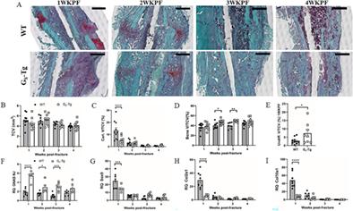
GαS, the stimulatory G protein α-subunit that raises intracellular cAMP levels by activating adenylyl cyclase, plays a vital role in bone development, maintenance, and remodeling. Previously, using transgenic mice overexpressing GαS in osteoblasts (GS-Tg), we demonstrated the influence of osteoblast GαS level on osteogenesis, bone turnover, and skeletal responses to hyperparathyroidism. To further investigate whether alterations in GαS levels affect endochondral bone repair, a postnatal bone regenerative process that recapitulates embryonic bone development, we performed stabilized tibial osteotomy in male GS-Tg mice at 8 weeks of age and examined the progression of fracture healing by micro-CT, histomorphometry, and gene expression analysis over a 4-week period. Bone fractures from GS-Tg mice exhibited diminished cartilage formation at the time of peak soft callus formation at 1 week post-fracture followed by significantly enhanced callus mineralization and new bone formation at 2 weeks post-fracture. The opposing effects on chondrogenesis and osteogenesis were validated by downregulation of chondrogenic markers and upregulation of osteogenic markers. Histomorphometric analysis at times of increased bone formation (2 and 3 weeks post-fracture) revealed excess fibroblast-like cells on newly formed woven bone surfaces and elevated osteocyte density in GS-Tg fractures. Coincident with enhanced callus mineralization and bone formation, GS-Tg mice showed elevated active β-catenin and Wntless proteins in osteoblasts at 2 weeks post-fracture, further substantiated by increased mRNA encoding various canonical Wnts and Wnt target genes, suggesting elevated osteoblastic Wnt secretion and Wnt/β-catenin signaling. The GS-Tg bony callus at 4 weeks post-fracture exhibited greater mineral density and decreased polar moment of inertia, resulting in improved material stiffness. These findings highlight that elevated GαS levels increase Wnt signaling, conferring an increased osteogenic differentiation potential at the expense of chondrogenic differentiation, resulting in improved mechanical integrity. © 2023 The Authors. JBMR Plus published by Wiley Periodicals LLC. on behalf of American Society for Bone and Mineral Research.

在小鼠骨折修复过程中,成骨细胞 GαS 的增加通过抑制软骨和增强胼胝体矿化促进骨化
GαS是刺激性G蛋白α亚基,通过激活腺苷酸环化酶提高细胞内cAMP水平,在骨骼发育、维持和重塑过程中发挥着重要作用。此前,我们利用在成骨细胞中过表达 GαS 的转基因小鼠(GS-Tg)证明了成骨细胞 GαS 水平对骨生成、骨转换和骨骼对甲状旁腺功能亢进反应的影响。为了进一步研究GαS水平的改变是否会影响软骨内骨修复(一种重现胚胎骨发育的产后骨再生过程),我们在雄性GS-Tg小鼠8周龄时对其进行了稳定的胫骨截骨术,并在4周时间内通过显微CT、组织形态学和基因表达分析检查了骨折愈合的进展。GS-Tg小鼠的骨折在骨折后1周达到软茧形成高峰时软骨形成减少,随后在骨折后2周胼胝矿化和新骨形成明显增强。软骨生成标志物的下调和成骨标志物的上调验证了对软骨生成和成骨的相反影响。在骨形成增加时(骨折后2周和3周)进行的组织形态学分析显示,GS-Tg骨折患者新形成的编织骨表面有过量的成纤维细胞,骨细胞密度升高。在胼胝体矿化和骨形成增强的同时,GS-Tg 小鼠在骨折后 2 周显示成骨细胞中活性 β-catenin 和 Wntless 蛋白升高,编码各种典型 Wnts 和 Wnt 靶基因的 mRNA 增加进一步证实了这一点,表明成骨细胞 Wnt 分泌和 Wnt/β-catenin 信号传导增强。骨折后4周的GS-Tg骨性胼胝体显示出更高的矿物质密度和更低的极性惯性矩,从而改善了材料的硬度。这些发现突出表明,GαS水平升高会增加Wnt信号传导,从而提高成骨分化潜能,牺牲软骨分化,从而改善机械完整性。© 2023 作者。JBMR Plus 由 Wiley Periodicals LLC.代表美国骨与矿物质研究学会出版。
本文章由计算机程序翻译,如有差异,请以英文原文为准。


 求助内容:
求助内容: 应助结果提醒方式:


