David Rutherford , Kateřina Kolářová , Jaroslav Čech , Petr Haušild , Jaroslav Kuliček , Egor Ukraintsev , Štěpán Stehlík , Radek Dao , Jan Neuman , Bohuslav Rezek
{"title":"Correlative atomic force microscopy and scanning electron microscopy of bacteria-diamond-metal nanocomposites","authors":"David Rutherford , Kateřina Kolářová , Jaroslav Čech , Petr Haušild , Jaroslav Kuliček , Egor Ukraintsev , Štěpán Stehlík , Radek Dao , Jan Neuman , Bohuslav Rezek","doi":"10.1016/j.ultramic.2023.113909","DOIUrl":null,"url":null,"abstract":"<div><p>Research investigating the interface between biological organisms and nanomaterials nowadays requires multi-faceted microscopic methods to elucidate the interaction mechanisms and effects. Here we describe a novel approach and methodology correlating data from an atomic force microscope inside a scanning electron microscope (AFM-in-SEM). This approach is demonstrated on bacteria-diamond-metal nanocomposite samples relevant in current life science research. We describe a procedure for preparing such multi-component test samples containing <em>E. coli</em> bacteria and chitosan-coated hydrogenated nanodiamonds decorated with silver nanoparticles on a carbon-coated gold grid. Microscopic topography information (AFM) is combined with chemical, material, and morphological information (SEM using SE and BSE at varied acceleration voltages) from the same region of interest and processed to create 3D correlative probe-electron microscopy (CPEM) images. We also establish a novel 3D RGB color image algorithm for merging multiple SE/BSE data from SEM with the AFM surface topography data which provides additional information about microscopic interaction of the diamond-metal nanocomposite with bacteria, not achievable by individual analyses. The methodology of CPEM data interpretation is independently corroborated by further <em>in-situ</em> (EDS) and <em>ex-situ</em> (micro-Raman) chemical characterization as well as by force volume AFM analysis. We also discuss the broader applicability and benefits of the methodology for life science research.</p></div>","PeriodicalId":23439,"journal":{"name":"Ultramicroscopy","volume":"258 ","pages":"Article 113909"},"PeriodicalIF":2.1000,"publicationDate":"2023-12-14","publicationTypes":"Journal Article","fieldsOfStudy":null,"isOpenAccess":false,"openAccessPdf":"","citationCount":"0","resultStr":null,"platform":"Semanticscholar","paperid":null,"PeriodicalName":"Ultramicroscopy","FirstCategoryId":"5","ListUrlMain":"https://www.sciencedirect.com/science/article/pii/S0304399123002267","RegionNum":3,"RegionCategory":"工程技术","ArticlePicture":[],"TitleCN":null,"AbstractTextCN":null,"PMCID":null,"EPubDate":"","PubModel":"","JCR":"Q2","JCRName":"MICROSCOPY","Score":null,"Total":0}
引用次数: 0
Abstract
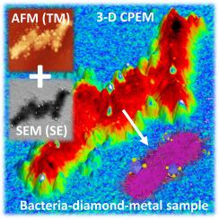
Research investigating the interface between biological organisms and nanomaterials nowadays requires multi-faceted microscopic methods to elucidate the interaction mechanisms and effects. Here we describe a novel approach and methodology correlating data from an atomic force microscope inside a scanning electron microscope (AFM-in-SEM). This approach is demonstrated on bacteria-diamond-metal nanocomposite samples relevant in current life science research. We describe a procedure for preparing such multi-component test samples containing E. coli bacteria and chitosan-coated hydrogenated nanodiamonds decorated with silver nanoparticles on a carbon-coated gold grid. Microscopic topography information (AFM) is combined with chemical, material, and morphological information (SEM using SE and BSE at varied acceleration voltages) from the same region of interest and processed to create 3D correlative probe-electron microscopy (CPEM) images. We also establish a novel 3D RGB color image algorithm for merging multiple SE/BSE data from SEM with the AFM surface topography data which provides additional information about microscopic interaction of the diamond-metal nanocomposite with bacteria, not achievable by individual analyses. The methodology of CPEM data interpretation is independently corroborated by further in-situ (EDS) and ex-situ (micro-Raman) chemical characterization as well as by force volume AFM analysis. We also discuss the broader applicability and benefits of the methodology for life science research.
期刊介绍:
Ultramicroscopy is an established journal that provides a forum for the publication of original research papers, invited reviews and rapid communications. The scope of Ultramicroscopy is to describe advances in instrumentation, methods and theory related to all modes of microscopical imaging, diffraction and spectroscopy in the life and physical sciences.


 求助内容:
求助内容: 应助结果提醒方式:


