Positional relationships of the origin and course of zygomaticus major with the nasal ala, tragus, philtrum, and lateral canthus for aesthetic treatments and surgeries
Hyun Jin Park, Jin Seo Park, Joe Iwanaga, R. Shane Tubbs, Mi-Sun Hur
{"title":"Positional relationships of the origin and course of zygomaticus major with the nasal ala, tragus, philtrum, and lateral canthus for aesthetic treatments and surgeries","authors":"Hyun Jin Park, Jin Seo Park, Joe Iwanaga, R. Shane Tubbs, Mi-Sun Hur","doi":"10.1007/s00276-023-03271-z","DOIUrl":null,"url":null,"abstract":"<h3 data-test=\"abstract-sub-heading\">Purpose</h3><p>The aim of this study was to characterize the origin and course of the zygomaticus major muscle (Zmj) with its topographic relationships with the nasal ala, tragus, philtrum, and lateral canthus.</p><h3 data-test=\"abstract-sub-heading\">Methods</h3><p>The Zmj was examined in 50 specimens of 25 embalmed adult Korean cadavers. Facial muscles were dissected to expose the origin and course of the Zmj in 48 specimens of 24 cadavers. The 25th cadaver was sectioned to obtain images of the Zmj.</p><h3 data-test=\"abstract-sub-heading\">Results</h3><p>The positional relationships of the Zmj origin with the nasal ala and the tragus were classified into three categories. A horizontal line through the center of the Zmj origin and the nasal ala passed through the tragus in 20 of 48 specimens (41.7%), the intertragic notch in 18 specimens (37.5%), and above the tragus in 10 specimens (20.8%). In a horizontal section of the head, the Zmj origin was located near the level of the nasal ala and tragus. In a coronal section of the head, the fibers of the Zmj arising at its origin were located close to the zygomatic bone, lateral to the zygomaticus minor muscle.</p><h3 data-test=\"abstract-sub-heading\">Conclusion</h3><p>By combining dissection with the analysis of sectioned images and ultrasound images of the Zmj, this study has yielded positional information for easily predicting the location of the origin and the course of the Zmj and its related structures underlying the skin.</p>","PeriodicalId":49296,"journal":{"name":"Surgical and Radiologic Anatomy","volume":"3 1","pages":""},"PeriodicalIF":1.2000,"publicationDate":"2023-12-13","publicationTypes":"Journal Article","fieldsOfStudy":null,"isOpenAccess":false,"openAccessPdf":"","citationCount":"0","resultStr":null,"platform":"Semanticscholar","paperid":null,"PeriodicalName":"Surgical and Radiologic Anatomy","FirstCategoryId":"3","ListUrlMain":"https://doi.org/10.1007/s00276-023-03271-z","RegionNum":4,"RegionCategory":"医学","ArticlePicture":[],"TitleCN":null,"AbstractTextCN":null,"PMCID":null,"EPubDate":"","PubModel":"","JCR":"Q3","JCRName":"ANATOMY & MORPHOLOGY","Score":null,"Total":0}
引用次数: 0
Abstract
Purpose
The aim of this study was to characterize the origin and course of the zygomaticus major muscle (Zmj) with its topographic relationships with the nasal ala, tragus, philtrum, and lateral canthus.
Methods
The Zmj was examined in 50 specimens of 25 embalmed adult Korean cadavers. Facial muscles were dissected to expose the origin and course of the Zmj in 48 specimens of 24 cadavers. The 25th cadaver was sectioned to obtain images of the Zmj.
Results
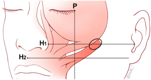
The positional relationships of the Zmj origin with the nasal ala and the tragus were classified into three categories. A horizontal line through the center of the Zmj origin and the nasal ala passed through the tragus in 20 of 48 specimens (41.7%), the intertragic notch in 18 specimens (37.5%), and above the tragus in 10 specimens (20.8%). In a horizontal section of the head, the Zmj origin was located near the level of the nasal ala and tragus. In a coronal section of the head, the fibers of the Zmj arising at its origin were located close to the zygomatic bone, lateral to the zygomaticus minor muscle.
Conclusion
By combining dissection with the analysis of sectioned images and ultrasound images of the Zmj, this study has yielded positional information for easily predicting the location of the origin and the course of the Zmj and its related structures underlying the skin.
期刊介绍:
Anatomy is a morphological science which cannot fail to interest the clinician. The practical application of anatomical research to clinical problems necessitates special adaptation and selectivity in choosing from numerous international works. Although there is a tendency to believe that meaningful advances in anatomy are unlikely, constant revision is necessary. Surgical and Radiologic Anatomy, the first international journal of Clinical anatomy has been created in this spirit.
Its goal is to serve clinicians, regardless of speciality-physicians, surgeons, radiologists or other specialists-as an indispensable aid with which they can improve their knowledge of anatomy. Each issue includes: Original papers, review articles, articles on the anatomical bases of medical, surgical and radiological techniques, articles of normal radiologic anatomy, brief reviews of anatomical publications of clinical interest.
Particular attention is given to high quality illustrations, which are indispensable for a better understanding of anatomical problems.
Surgical and Radiologic Anatomy is a journal written by anatomists for clinicians with a special interest in anatomy.

 求助内容:
求助内容: 应助结果提醒方式:


