Francesco Martino , Gennaro Ilardi , Silvia Varricchio , Daniela Russo , Rosa Maria Di Crescenzo , Stefania Staibano , Francesco Merolla
{"title":"A deep learning model to predict Ki-67 positivity in oral squamous cell carcinoma","authors":"Francesco Martino , Gennaro Ilardi , Silvia Varricchio , Daniela Russo , Rosa Maria Di Crescenzo , Stefania Staibano , Francesco Merolla","doi":"10.1016/j.jpi.2023.100354","DOIUrl":null,"url":null,"abstract":"<div><p>Anatomical pathology is undergoing its third revolution, transitioning from analogical to digital pathology and incorporating new artificial intelligence technologies into clinical practice. Aside from classification, detection, and segmentation models, predictive models are gaining traction since they can impact diagnostic processes and laboratory activity, lowering consumable usage and turnaround time. Our research aimed to create a deep-learning model to generate synthetic Ki-67 immunohistochemistry from Haematoxylin and Eosin (H&E) stained images. We used 175 oral squamous cell carcinoma (OSCC) from the University Federico II’s Pathology Unit’s archives to train our model to generate 4 Tissue Micro Arrays (TMAs). We sectioned one slide from each TMA, first stained with H&E and then re-stained with anti-Ki-67 immunohistochemistry (IHC). In digitised slides, cores were disarrayed, and the matching cores of the 2 stained were aligned to construct a dataset to train a Pix2Pix algorithm to convert H&E images to IHC. Pathologists could recognise the synthetic images in only half of the cases in a specially designed likelihood test. Hence, our model produced realistic synthetic images. We next used QuPath to quantify IHC positivity, achieving remarkable levels of agreement between genuine and synthetic IHC.</p><p>Furthermore, a categorical analysis employing 3 Ki-67 positivity cut-offs (5%, 10%, and 15%) revealed high positive-predictive values. Our model is a promising tool for collecting Ki-67 positivity information directly on H&E slides, reducing laboratory demand and improving patient management. It is also a valuable option for smaller laboratories to easily and quickly screen bioptic samples and prioritise them in a digital pathology workflow.</p></div>","PeriodicalId":37769,"journal":{"name":"Journal of Pathology Informatics","volume":"15 ","pages":"Article 100354"},"PeriodicalIF":0.0000,"publicationDate":"2023-11-22","publicationTypes":"Journal Article","fieldsOfStudy":null,"isOpenAccess":false,"openAccessPdf":"https://www.sciencedirect.com/science/article/pii/S2153353923001682/pdfft?md5=1e50c2def78451a443dc9e3c3d022cbc&pid=1-s2.0-S2153353923001682-main.pdf","citationCount":"0","resultStr":null,"platform":"Semanticscholar","paperid":null,"PeriodicalName":"Journal of Pathology Informatics","FirstCategoryId":"1085","ListUrlMain":"https://www.sciencedirect.com/science/article/pii/S2153353923001682","RegionNum":0,"RegionCategory":null,"ArticlePicture":[],"TitleCN":null,"AbstractTextCN":null,"PMCID":null,"EPubDate":"","PubModel":"","JCR":"Q2","JCRName":"Medicine","Score":null,"Total":0}
引用次数: 0
Abstract
Anatomical pathology is undergoing its third revolution, transitioning from analogical to digital pathology and incorporating new artificial intelligence technologies into clinical practice. Aside from classification, detection, and segmentation models, predictive models are gaining traction since they can impact diagnostic processes and laboratory activity, lowering consumable usage and turnaround time. Our research aimed to create a deep-learning model to generate synthetic Ki-67 immunohistochemistry from Haematoxylin and Eosin (H&E) stained images. We used 175 oral squamous cell carcinoma (OSCC) from the University Federico II’s Pathology Unit’s archives to train our model to generate 4 Tissue Micro Arrays (TMAs). We sectioned one slide from each TMA, first stained with H&E and then re-stained with anti-Ki-67 immunohistochemistry (IHC). In digitised slides, cores were disarrayed, and the matching cores of the 2 stained were aligned to construct a dataset to train a Pix2Pix algorithm to convert H&E images to IHC. Pathologists could recognise the synthetic images in only half of the cases in a specially designed likelihood test. Hence, our model produced realistic synthetic images. We next used QuPath to quantify IHC positivity, achieving remarkable levels of agreement between genuine and synthetic IHC.
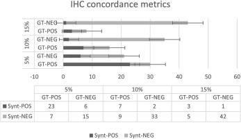
Furthermore, a categorical analysis employing 3 Ki-67 positivity cut-offs (5%, 10%, and 15%) revealed high positive-predictive values. Our model is a promising tool for collecting Ki-67 positivity information directly on H&E slides, reducing laboratory demand and improving patient management. It is also a valuable option for smaller laboratories to easily and quickly screen bioptic samples and prioritise them in a digital pathology workflow.
期刊介绍:
The Journal of Pathology Informatics (JPI) is an open access peer-reviewed journal dedicated to the advancement of pathology informatics. This is the official journal of the Association for Pathology Informatics (API). The journal aims to publish broadly about pathology informatics and freely disseminate all articles worldwide. This journal is of interest to pathologists, informaticians, academics, researchers, health IT specialists, information officers, IT staff, vendors, and anyone with an interest in informatics. We encourage submissions from anyone with an interest in the field of pathology informatics. We publish all types of papers related to pathology informatics including original research articles, technical notes, reviews, viewpoints, commentaries, editorials, symposia, meeting abstracts, book reviews, and correspondence to the editors. All submissions are subject to rigorous peer review by the well-regarded editorial board and by expert referees in appropriate specialties.

 求助内容:
求助内容: 应助结果提醒方式:


