Maria Antoniou, Dimitra Tsounidi, Panagiota S. Petrou, Konstantinos G. Beltsios, Sotirios E. Kakabakos
{"title":"Functionalization of silicon dioxide and silicon nitride surfaces with aminosilanes for optical biosensing applications","authors":"Maria Antoniou, Dimitra Tsounidi, Panagiota S. Petrou, Konstantinos G. Beltsios, Sotirios E. Kakabakos","doi":"10.1002/mds3.10072","DOIUrl":null,"url":null,"abstract":"<p>The development of optical biosensors based on silicon dioxide or silicon nitride transducers requires the chemical activation of their surface to achieve stable, repeatable and homogeneous binding of biomolecules. In the present study, the chemical activation of silicon dioxide and silicon nitride surfaces with 3-aminopropyl-triethoxysilane (APTES) was optimized so as to enable the immobilization of biomolecules by adsorption or covalent bonding. Chemical activation was performed with either aqueous or organic solution of APTES, and the surfaces were used to immobilize directly protein molecules by physical adsorption or further modified with glutaraldehyde to allow covalent binding of protein molecules. The protein immobilization capacity of the chemically activated silicon dioxide and silicon nitride surfaces was evaluated through incubation with mouse γ-globulins and reaction with a fluorescently labelled goat antimouse IgG antibody. By determining the surface fluorescence signal intensity, it was found that modification with 5% (v/v) APTES solution in ethanol followed by modification with glutaraldehyde provided 30% higher fluorescence signals than all the other protocols tested. In addition, this method provided the lower signal variation between different chips. To test the possible advantages of the chemical activation protocols for optical biosensing applications, they were also applied to a label-free white light interference spectroscopy sensor and evaluated through (a) real-time monitoring of the reaction between immobilized on the sensor surface mouse γ-globulins with an unlabelled goat antimouse IgG antibody and (b) a non-competitive immunoassay for the determination of C-reactive protein. The results showed that in case of antibody, physical absorption provided marginally higher binding capacity to covalent bonding.</p>","PeriodicalId":87324,"journal":{"name":"Medical devices & sensors","volume":"3 5","pages":""},"PeriodicalIF":0.0000,"publicationDate":"2020-03-12","publicationTypes":"Journal Article","fieldsOfStudy":null,"isOpenAccess":false,"openAccessPdf":"https://sci-hub-pdf.com/10.1002/mds3.10072","citationCount":"14","resultStr":null,"platform":"Semanticscholar","paperid":null,"PeriodicalName":"Medical devices & sensors","FirstCategoryId":"1085","ListUrlMain":"https://onlinelibrary.wiley.com/doi/10.1002/mds3.10072","RegionNum":0,"RegionCategory":null,"ArticlePicture":[],"TitleCN":null,"AbstractTextCN":null,"PMCID":null,"EPubDate":"","PubModel":"","JCR":"","JCRName":"","Score":null,"Total":0}
引用次数: 14
Abstract
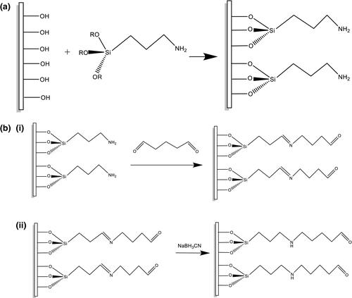
The development of optical biosensors based on silicon dioxide or silicon nitride transducers requires the chemical activation of their surface to achieve stable, repeatable and homogeneous binding of biomolecules. In the present study, the chemical activation of silicon dioxide and silicon nitride surfaces with 3-aminopropyl-triethoxysilane (APTES) was optimized so as to enable the immobilization of biomolecules by adsorption or covalent bonding. Chemical activation was performed with either aqueous or organic solution of APTES, and the surfaces were used to immobilize directly protein molecules by physical adsorption or further modified with glutaraldehyde to allow covalent binding of protein molecules. The protein immobilization capacity of the chemically activated silicon dioxide and silicon nitride surfaces was evaluated through incubation with mouse γ-globulins and reaction with a fluorescently labelled goat antimouse IgG antibody. By determining the surface fluorescence signal intensity, it was found that modification with 5% (v/v) APTES solution in ethanol followed by modification with glutaraldehyde provided 30% higher fluorescence signals than all the other protocols tested. In addition, this method provided the lower signal variation between different chips. To test the possible advantages of the chemical activation protocols for optical biosensing applications, they were also applied to a label-free white light interference spectroscopy sensor and evaluated through (a) real-time monitoring of the reaction between immobilized on the sensor surface mouse γ-globulins with an unlabelled goat antimouse IgG antibody and (b) a non-competitive immunoassay for the determination of C-reactive protein. The results showed that in case of antibody, physical absorption provided marginally higher binding capacity to covalent bonding.

 求助内容:
求助内容: 应助结果提醒方式:


