Decreased 5‐HT1A binding in mild Alzheimer's disease—A positron emission tomography study
引用次数: 0
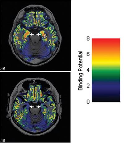
Abstract
Decreased 5‐HT1A receptor binding has been associated with Alzheimer's disease (AD) and interpreted as a consequence of neuron loss. The purpose of the present study was to compare [11C]WAY100635 binding to the 5‐HT1A receptor in the hippocampus, entorhinal cortex, amygdala and pericalcarine cortex in mild AD patients and elderly controls. AD patients (n = 7) and elderly control subjects (n = 8) were examined with positron emission tomography (PET) and [11C]WAY100635. PET data acquisition was performed with an ECAT EXACT HR system. Wavelet‐aided parametric images of nondisplaceable binding potential (BPND) were generated using Logan's graphical analysis with cerebellum as the reference region. Correction for partial volume effects was performed with the Müller–Gärtner method. Regions of interest (ROIs) were applied to the individual parametric images, and the regional BPND was calculated as the average parametric voxel value within each ROI. In addition to comparisons between subject groups, correlations between BPND values and scores on the Mini‐Mental State Examination, Disability Assessment for Dementia (DAD), and Neuropsychiatric Inventory were expressed by Pearson correlation coefficients. Mean regional BPND was lower in AD patients than in control subjects, and the difference was statistically significant for the hippocampus, entorhinal cortex, and amygdala. A statistically significant correlation was obtained between hippocampal BPND values and DAD scores. The results of the present study corroborate and extend previous findings of decreased 5‐HT1A binding in AD and strengthen the support for 5‐HT1A receptor PET as a tool for the assessment of neurodegenerative changes in mild AD.
轻度阿尔茨海默病中5‐HT1A结合减少——正电子发射断层扫描研究
5‐HT1A受体结合减少与阿尔茨海默病(AD)有关,并被解释为神经元丢失的结果。本研究的目的是比较[11C]WAY100635在轻度AD患者和老年对照中与海马体、内嗅皮层、杏仁核和骨膜外皮层中与5‐HT1A受体的结合情况。采用正电子发射断层扫描(PET)和[11C]WAY100635对AD患者(n = 7)和老年对照(n = 8)进行检查。PET数据采集采用ECAT EXACT HR系统。以小脑为参考区域,采用Logan图形分析方法生成了小波辅助的不可置换结合电位(BPND)参数图像。使用Müller-Gärtner方法对部分体积效应进行校正。将感兴趣区域(ROI)应用于单个参数图像,并计算区域BPND作为每个ROI内的平均参数体素值。除了受试者组之间的比较外,BPND值与迷你精神状态检查、痴呆残疾评估(DAD)和神经精神量表得分之间的相关性通过Pearson相关系数表示。AD患者的平均区域BPND低于对照组,海马、内嗅皮层和杏仁核的差异有统计学意义。海马BPND值与DAD评分之间有统计学意义的相关性。本研究的结果证实并扩展了先前关于AD中5‐HT1A结合减少的发现,并加强了对5‐HT1A受体PET作为轻度AD神经退行性改变评估工具的支持。
本文章由计算机程序翻译,如有差异,请以英文原文为准。
求助全文
约1分钟内获得全文
求助全文

 求助内容:
求助内容: 应助结果提醒方式:


