S. Gomez-Pena , A. Rueda de Eusebio , J. Arrazola García , P. Romero Fernández , M.J. Moreno Casado , A.M. Crespo Rodríguez
{"title":"Actualización de los tumores cartilaginosos según la clasificación de la OMS de 2020","authors":"S. Gomez-Pena , A. Rueda de Eusebio , J. Arrazola García , P. Romero Fernández , M.J. Moreno Casado , A.M. Crespo Rodríguez","doi":"10.1016/j.rx.2023.05.003","DOIUrl":null,"url":null,"abstract":"<div><p>Cartilaginous tumours are a large and heterogeneous group of neoplasms characterised by the presence of a chondroid matrix, with lobular growth and arcuate, ring-like or popcorn-like calcification patterns. MRI shows hyperintensity in T2-weighted sequences and a lobulated or septal relief in postcontrast images.</p><p>In the WHO 2020 classification, chondral tumours are classified as benign, intermediate or malignant. Despite technological advances, they continue to pose a challenge for both the radiologist and the pathologist, being the main difficulty the differentiation between benign and malignant tumours, which is why they require a multidisciplinary approach.</p><p>This paper describes the main changes introduced in the 2020 update, describes the imaging characteristics of the main cartilaginous tumours and provides the radiological keys to differentiate between benign and malignant tumours.</p></div>","PeriodicalId":31509,"journal":{"name":"RADIOLOGIA","volume":"66 1","pages":"Pages 57-69"},"PeriodicalIF":1.1000,"publicationDate":"2024-01-01","publicationTypes":"Journal Article","fieldsOfStudy":null,"isOpenAccess":false,"openAccessPdf":"","citationCount":"0","resultStr":null,"platform":"Semanticscholar","paperid":null,"PeriodicalName":"RADIOLOGIA","FirstCategoryId":"1085","ListUrlMain":"https://www.sciencedirect.com/science/article/pii/S0033833823000978","RegionNum":0,"RegionCategory":null,"ArticlePicture":[],"TitleCN":null,"AbstractTextCN":null,"PMCID":null,"EPubDate":"","PubModel":"","JCR":"Q3","JCRName":"RADIOLOGY, NUCLEAR MEDICINE & MEDICAL IMAGING","Score":null,"Total":0}
引用次数: 0
Abstract
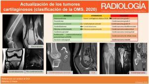
Cartilaginous tumours are a large and heterogeneous group of neoplasms characterised by the presence of a chondroid matrix, with lobular growth and arcuate, ring-like or popcorn-like calcification patterns. MRI shows hyperintensity in T2-weighted sequences and a lobulated or septal relief in postcontrast images.
In the WHO 2020 classification, chondral tumours are classified as benign, intermediate or malignant. Despite technological advances, they continue to pose a challenge for both the radiologist and the pathologist, being the main difficulty the differentiation between benign and malignant tumours, which is why they require a multidisciplinary approach.
This paper describes the main changes introduced in the 2020 update, describes the imaging characteristics of the main cartilaginous tumours and provides the radiological keys to differentiate between benign and malignant tumours.
RADIOLOGIARADIOLOGY, NUCLEAR MEDICINE & MEDICAL IMAGING-
CiteScore
1.60
自引率
7.70%
发文量
105
审稿时长
52 days
期刊介绍:
La mejor revista para conocer de primera mano los originales más relevantes en la especialidad y las revisiones, casos y notas clínicas de mayor interés profesional. Además es la Publicación Oficial de la Sociedad Española de Radiología Médica.
 求助内容:
求助内容: 应助结果提醒方式:


