Radiomics Analysis of Gray-Scale Ultrasonographic Images of Papillary Thyroid Carcinoma > 1 cm: Potential Biomarker for the Prediction of Lymph Node Metastasis.
Hyun Jung Chung, Kyunghwa Han, Eunjung Lee, Jung Hyun Yoon, Vivian Youngjean Park, Mina Lee, Eun Cho, Jin Young Kwak
{"title":"Radiomics Analysis of Gray-Scale Ultrasonographic Images of Papillary Thyroid Carcinoma > 1 cm: Potential Biomarker for the Prediction of Lymph Node Metastasis.","authors":"Hyun Jung Chung, Kyunghwa Han, Eunjung Lee, Jung Hyun Yoon, Vivian Youngjean Park, Mina Lee, Eun Cho, Jin Young Kwak","doi":"10.3348/jksr.2021.0155","DOIUrl":null,"url":null,"abstract":"<p><strong>Purpose: </strong>This study aimed to investigate radiomics analysis of ultrasonographic images to develop a potential biomarker for predicting lymph node metastasis in papillary thyroid carcinoma (PTC) patients.</p><p><strong>Materials and methods: </strong>This study included 431 PTC patients from August 2013 to May 2014 and classified them into the training and validation sets. A total of 730 radiomics features, including texture matrices of gray-level co-occurrence matrix and gray-level run-length matrix and single-level discrete two-dimensional wavelet transform and other functions, were obtained. The least absolute shrinkage and selection operator method was used for selecting the most predictive features in the training data set.</p><p><strong>Results: </strong>Lymph node metastasis was associated with the radiomics score (<i>p</i> < 0.001). It was also associated with other clinical variables such as young age (<i>p</i> = 0.007) and large tumor size (<i>p</i> = 0.007). The area under the receiver operating characteristic curve was 0.687 (95% confidence interval: 0.616-0.759) for the training set and 0.650 (95% confidence interval: 0.575-0.726) for the validation set.</p><p><strong>Conclusion: </strong>This study showed the potential of ultrasonography-based radiomics to predict cervical lymph node metastasis in patients with PTC; thus, ultrasonography-based radiomics can act as a biomarker for PTC.</p>","PeriodicalId":17455,"journal":{"name":"Journal of the Korean Society of Radiology","volume":"84 1","pages":"185-196"},"PeriodicalIF":0.0000,"publicationDate":"2023-01-01","publicationTypes":"Journal Article","fieldsOfStudy":null,"isOpenAccess":false,"openAccessPdf":"https://ftp.ncbi.nlm.nih.gov/pub/pmc/oa_pdf/15/b0/jksr-84-185.PMC9935950.pdf","citationCount":"0","resultStr":null,"platform":"Semanticscholar","paperid":null,"PeriodicalName":"Journal of the Korean Society of Radiology","FirstCategoryId":"1085","ListUrlMain":"https://doi.org/10.3348/jksr.2021.0155","RegionNum":0,"RegionCategory":null,"ArticlePicture":[],"TitleCN":null,"AbstractTextCN":null,"PMCID":null,"EPubDate":"","PubModel":"","JCR":"Q4","JCRName":"Medicine","Score":null,"Total":0}
引用次数: 0
Abstract
Purpose: This study aimed to investigate radiomics analysis of ultrasonographic images to develop a potential biomarker for predicting lymph node metastasis in papillary thyroid carcinoma (PTC) patients.
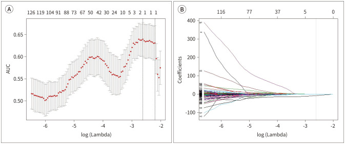
Materials and methods: This study included 431 PTC patients from August 2013 to May 2014 and classified them into the training and validation sets. A total of 730 radiomics features, including texture matrices of gray-level co-occurrence matrix and gray-level run-length matrix and single-level discrete two-dimensional wavelet transform and other functions, were obtained. The least absolute shrinkage and selection operator method was used for selecting the most predictive features in the training data set.
Results: Lymph node metastasis was associated with the radiomics score (p < 0.001). It was also associated with other clinical variables such as young age (p = 0.007) and large tumor size (p = 0.007). The area under the receiver operating characteristic curve was 0.687 (95% confidence interval: 0.616-0.759) for the training set and 0.650 (95% confidence interval: 0.575-0.726) for the validation set.
Conclusion: This study showed the potential of ultrasonography-based radiomics to predict cervical lymph node metastasis in patients with PTC; thus, ultrasonography-based radiomics can act as a biomarker for PTC.

 求助内容:
求助内容: 应助结果提醒方式:


