Kurt G Schilling, Muwei Li, Francois Rheault, Zhaohua Ding, Adam W Anderson, Hakmook Kang, Bennett A Landman, John C Gore
{"title":"Anomalous and heterogeneous characteristics of the BOLD hemodynamic response function in white matter.","authors":"Kurt G Schilling, Muwei Li, Francois Rheault, Zhaohua Ding, Adam W Anderson, Hakmook Kang, Bennett A Landman, John C Gore","doi":"10.1093/texcom/tgac035","DOIUrl":null,"url":null,"abstract":"<p><p>Detailed knowledge of the BOLD hemodynamic response function (HRF) is crucial for accurate analyses and interpretation of functional MRI data. Considerable efforts have been made to characterize the HRF in gray matter (GM), but much less attention has been paid to BOLD effects in white matter (WM). However, several recent reports have demonstrated reliable detection and analyses of WM BOLD signals both after stimulation and in a resting state. WM and GM differ in composition, energy requirements, and blood flow, so their neurovascular couplings also may well be different. We aimed to derive a comprehensive characterization of the HRF in WM across a population, including accurate measurements of its shape and its variation along and between WM pathways, using resting-state fMRI acquisitions. Our results show that the HRF is significantly different between WM and GM. Features of the HRF, such as a prominent initial dip, show strong relationships with features of the tissue microstructure derived from diffusion imaging, and these relationships differ between WM and GM, consistent with BOLD signal fluctuations reflecting different energy demands and neurovascular couplings in tissues of different composition and function. We also show that the HRF varies in shape significantly along WM pathways and is different between different WM pathways, suggesting the temporal evolution of BOLD signals after an event vary in different parts of the WM. These features of the HRF in WM are especially relevant for interpretation of the biophysical basis of BOLD effects in WM.</p>","PeriodicalId":72551,"journal":{"name":"Cerebral cortex communications","volume":"3 3","pages":"tgac035"},"PeriodicalIF":0.0000,"publicationDate":"2022-01-01","publicationTypes":"Journal Article","fieldsOfStudy":null,"isOpenAccess":false,"openAccessPdf":"https://www.ncbi.nlm.nih.gov/pmc/articles/PMC9519945/pdf/","citationCount":"8","resultStr":null,"platform":"Semanticscholar","paperid":null,"PeriodicalName":"Cerebral cortex communications","FirstCategoryId":"1085","ListUrlMain":"https://doi.org/10.1093/texcom/tgac035","RegionNum":0,"RegionCategory":null,"ArticlePicture":[],"TitleCN":null,"AbstractTextCN":null,"PMCID":null,"EPubDate":"","PubModel":"","JCR":"","JCRName":"","Score":null,"Total":0}
引用次数: 8
Abstract
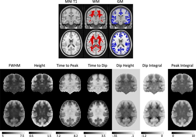
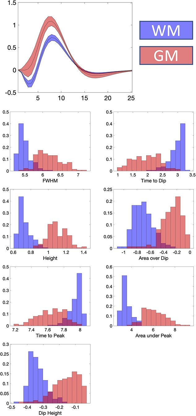
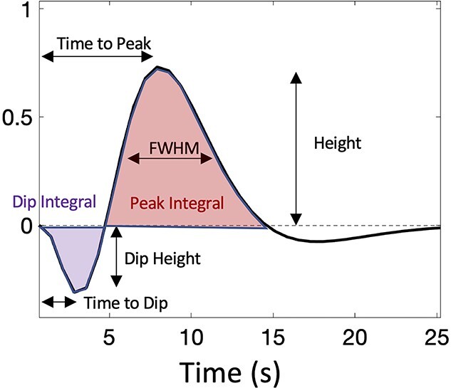
Detailed knowledge of the BOLD hemodynamic response function (HRF) is crucial for accurate analyses and interpretation of functional MRI data. Considerable efforts have been made to characterize the HRF in gray matter (GM), but much less attention has been paid to BOLD effects in white matter (WM). However, several recent reports have demonstrated reliable detection and analyses of WM BOLD signals both after stimulation and in a resting state. WM and GM differ in composition, energy requirements, and blood flow, so their neurovascular couplings also may well be different. We aimed to derive a comprehensive characterization of the HRF in WM across a population, including accurate measurements of its shape and its variation along and between WM pathways, using resting-state fMRI acquisitions. Our results show that the HRF is significantly different between WM and GM. Features of the HRF, such as a prominent initial dip, show strong relationships with features of the tissue microstructure derived from diffusion imaging, and these relationships differ between WM and GM, consistent with BOLD signal fluctuations reflecting different energy demands and neurovascular couplings in tissues of different composition and function. We also show that the HRF varies in shape significantly along WM pathways and is different between different WM pathways, suggesting the temporal evolution of BOLD signals after an event vary in different parts of the WM. These features of the HRF in WM are especially relevant for interpretation of the biophysical basis of BOLD effects in WM.

 求助内容:
求助内容: 应助结果提醒方式:


