{"title":"Ruptured Basilar Artery Perforator Aneurysm Definitely Diagnosed with Intraoperative Microsurgical Findings: Case Report and Literature Review.","authors":"Takahiro Kumagawa, Naoki Otani, Yuzo Kakei, Hiroshi Negishi, Takeshi Suma, Atsuo Yoshino","doi":"10.2176/jns-nmc.2022-0184","DOIUrl":null,"url":null,"abstract":"<p><p>Initial three-dimensional computed tomography and cerebral angiography fail to identify any aneurysm in 20% of cases of subarachnoid hemorrhage. Basilar artery (BA) perforator aneurysms are rare, and approximately 30%-60% were not identified by initial angiography. A 71-year-old male was transferred with a sudden onset of headache and loss of consciousness. Computed tomography demonstrated subarachnoid hemorrhage, but no ruptured aneurysm was detected. Repeat preoperative cerebral angiography indicated a bifurcation aneurysm of the circumflex branch of the superior cerebellar artery perforator, but microsurgical observation identified the BA perforator aneurysm. If the location of the BA perforator aneurysm cannot be clearly identified, as in this case, repeat angiography should be considered, and the treatment strategy should be decided based on a detailed consideration of the site of the aneurysm.</p>","PeriodicalId":19260,"journal":{"name":"NMC Case Report Journal","volume":"10 ","pages":"1-7"},"PeriodicalIF":0.0000,"publicationDate":"2023-01-01","publicationTypes":"Journal Article","fieldsOfStudy":null,"isOpenAccess":false,"openAccessPdf":"https://ftp.ncbi.nlm.nih.gov/pub/pmc/oa_pdf/0f/a7/2188-4226-10-0001.PMC9894615.pdf","citationCount":"0","resultStr":null,"platform":"Semanticscholar","paperid":null,"PeriodicalName":"NMC Case Report Journal","FirstCategoryId":"1085","ListUrlMain":"https://doi.org/10.2176/jns-nmc.2022-0184","RegionNum":0,"RegionCategory":null,"ArticlePicture":[],"TitleCN":null,"AbstractTextCN":null,"PMCID":null,"EPubDate":"","PubModel":"","JCR":"","JCRName":"","Score":null,"Total":0}
引用次数: 0
Abstract
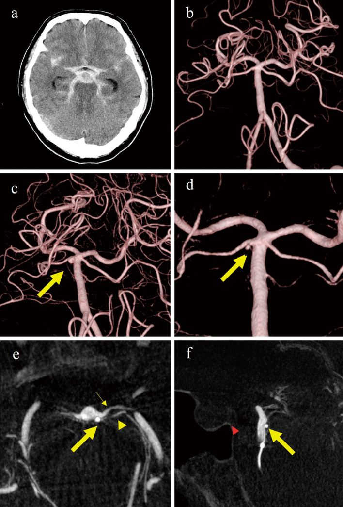
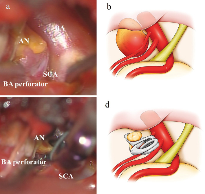
Initial three-dimensional computed tomography and cerebral angiography fail to identify any aneurysm in 20% of cases of subarachnoid hemorrhage. Basilar artery (BA) perforator aneurysms are rare, and approximately 30%-60% were not identified by initial angiography. A 71-year-old male was transferred with a sudden onset of headache and loss of consciousness. Computed tomography demonstrated subarachnoid hemorrhage, but no ruptured aneurysm was detected. Repeat preoperative cerebral angiography indicated a bifurcation aneurysm of the circumflex branch of the superior cerebellar artery perforator, but microsurgical observation identified the BA perforator aneurysm. If the location of the BA perforator aneurysm cannot be clearly identified, as in this case, repeat angiography should be considered, and the treatment strategy should be decided based on a detailed consideration of the site of the aneurysm.

 求助内容:
求助内容: 应助结果提醒方式:


