Maria Cuadrado-Vilanova, Victor Burgueño, Leire Balaguer-Lluna, Rosario Aschero, Helena Castillo-Ecija, Jing Liu, Sara Perez-Jaume, Guillem Pascual-Pasto, Nagore G Olaciregui, Soledad Gomez-Gonzalez, Genoveva Correa, Mariona Suñol, Paula Schaiquevich, François Radvanyi, Cinzia Lavarino, Jaume Mora, Jaume Catala-Mora, Guillermo L Chantada, Angel M Carcaboso
{"title":"Follow-up of intraocular retinoblastoma through the quantitative analysis of conserved nuclear DNA sequences in aqueous humor from patients","authors":"Maria Cuadrado-Vilanova, Victor Burgueño, Leire Balaguer-Lluna, Rosario Aschero, Helena Castillo-Ecija, Jing Liu, Sara Perez-Jaume, Guillem Pascual-Pasto, Nagore G Olaciregui, Soledad Gomez-Gonzalez, Genoveva Correa, Mariona Suñol, Paula Schaiquevich, François Radvanyi, Cinzia Lavarino, Jaume Mora, Jaume Catala-Mora, Guillermo L Chantada, Angel M Carcaboso","doi":"10.1002/cjp2.296","DOIUrl":null,"url":null,"abstract":"<p>Fundoscopy is the standard method for diagnosis and follow-up of intraocular retinoblastoma, but it is sometimes insufficient to discern whether tumors are inactivated following treatments. In this work, we hypothesized that the amount of conserved nuclear DNA sequences in the cell-free DNA (cfDNA) fraction of the aqueous humor (AH) might complement fundoscopy for retinoblastoma follow-up. To address our hypothesis, we developed highly sensitive droplet digital polymerase chain reaction (ddPCR) methods to quantify highly conserved DNA sequences of nucleus-encoded genes (<i>GAPDH</i> and <i>B4GALNT1)</i> and of a mitochondrial gene, <i>MT-ATP6</i>. We obtained AH samples during intravitreal treatments. We analyzed 42 AH samples from 25 patients with intraocular retinoblastoma and 11 AH from controls (non-cancer patients). According to clinical criteria, we grouped patients as having progression-free or progressive retinoblastoma. cfDNA concentration in the AH was similar in both retinoblastoma groups. Copy counts for nucleus-derived sequences of <i>GAPDH</i> and <i>B4GALNT1</i> were significantly higher in the AH from patients with progressive disease, compared to the AH from progression-free patients and control non-cancer patients. The presence of mitochondrial DNA in the AH explained that both retinoblastoma groups had similar cfDNA concentration in AH. The optimal cut-off point for discriminating between progressive and progression-free retinoblastomas was 108 <i>GAPDH</i> copies per reaction. Among patients having serial AH samples analyzed during their intravitreal chemotherapy, <i>GAPDH</i> copies were high and decreased below the cut-off point in those patients responding to chemotherapy. In contrast, one non-responder patient remained with values above the cut-off during follow-up, until enucleation. We conclude that the measurement of conserved nuclear gene sequences in AH allows follow-up of intraocular retinoblastoma during intravitreal treatment. The method is applicable to all patients and could be relevant for those in which fundoscopy evaluation is inconclusive.</p>","PeriodicalId":48612,"journal":{"name":"Journal of Pathology Clinical Research","volume":null,"pages":null},"PeriodicalIF":3.4000,"publicationDate":"2022-09-23","publicationTypes":"Journal Article","fieldsOfStudy":null,"isOpenAccess":false,"openAccessPdf":"https://pathsocjournals.onlinelibrary.wiley.com/doi/epdf/10.1002/cjp2.296","citationCount":"3","resultStr":null,"platform":"Semanticscholar","paperid":null,"PeriodicalName":"Journal of Pathology Clinical Research","FirstCategoryId":"3","ListUrlMain":"https://onlinelibrary.wiley.com/doi/10.1002/cjp2.296","RegionNum":2,"RegionCategory":"医学","ArticlePicture":[],"TitleCN":null,"AbstractTextCN":null,"PMCID":null,"EPubDate":"","PubModel":"","JCR":"Q1","JCRName":"PATHOLOGY","Score":null,"Total":0}
引用次数: 3
Abstract
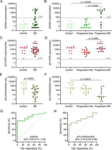
Fundoscopy is the standard method for diagnosis and follow-up of intraocular retinoblastoma, but it is sometimes insufficient to discern whether tumors are inactivated following treatments. In this work, we hypothesized that the amount of conserved nuclear DNA sequences in the cell-free DNA (cfDNA) fraction of the aqueous humor (AH) might complement fundoscopy for retinoblastoma follow-up. To address our hypothesis, we developed highly sensitive droplet digital polymerase chain reaction (ddPCR) methods to quantify highly conserved DNA sequences of nucleus-encoded genes (GAPDH and B4GALNT1) and of a mitochondrial gene, MT-ATP6. We obtained AH samples during intravitreal treatments. We analyzed 42 AH samples from 25 patients with intraocular retinoblastoma and 11 AH from controls (non-cancer patients). According to clinical criteria, we grouped patients as having progression-free or progressive retinoblastoma. cfDNA concentration in the AH was similar in both retinoblastoma groups. Copy counts for nucleus-derived sequences of GAPDH and B4GALNT1 were significantly higher in the AH from patients with progressive disease, compared to the AH from progression-free patients and control non-cancer patients. The presence of mitochondrial DNA in the AH explained that both retinoblastoma groups had similar cfDNA concentration in AH. The optimal cut-off point for discriminating between progressive and progression-free retinoblastomas was 108 GAPDH copies per reaction. Among patients having serial AH samples analyzed during their intravitreal chemotherapy, GAPDH copies were high and decreased below the cut-off point in those patients responding to chemotherapy. In contrast, one non-responder patient remained with values above the cut-off during follow-up, until enucleation. We conclude that the measurement of conserved nuclear gene sequences in AH allows follow-up of intraocular retinoblastoma during intravitreal treatment. The method is applicable to all patients and could be relevant for those in which fundoscopy evaluation is inconclusive.
期刊介绍:
The Journal of Pathology: Clinical Research and The Journal of Pathology serve as translational bridges between basic biomedical science and clinical medicine with particular emphasis on, but not restricted to, tissue based studies.
The focus of The Journal of Pathology: Clinical Research is the publication of studies that illuminate the clinical relevance of research in the broad area of the study of disease. Appropriately powered and validated studies with novel diagnostic, prognostic and predictive significance, and biomarker discover and validation, will be welcomed. Studies with a predominantly mechanistic basis will be more appropriate for the companion Journal of Pathology.
 求助内容:
求助内容: 应助结果提醒方式:


