Gastrointestinal Metastatic Melanoma: The Key for Diagnosis.
IF 1
Q4 GASTROENTEROLOGY & HEPATOLOGY
引用次数: 2
Abstract
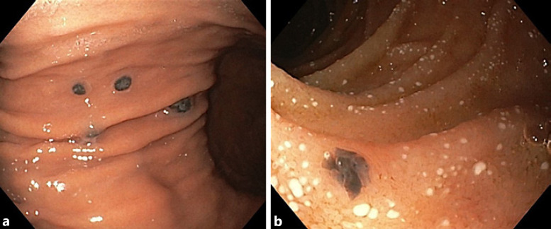
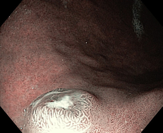
An 80-year-old Caucasian woman was hospitalized with a 2-month course of intermittent fever (max. 38 ° C), asthenia, weight loss (12%), anorexia and nausea. Her medical history includes breast cancer submitted to radical mastectomy and axillary lymph node dissection, papillary thyroid carcinoma and pulmonary and ocular tuberculosis that had been treated more than 5 years previously. She had heart failure, arterial hypertension, dyslipidaemia and obesity under treatment. Physical examination showed obesity and left upper limb lymphedema. Abdominal and rectal examinations were unremarkable. A laboratory study revealed iron deficiency anaemia with haemoglobin 10 g/dL and ferritin 10 ng/mL (normal = 10–120 ng/mL), elevated lactate dehydrogenase 1,379 U/L (normal <247 U/L), aspartate transaminase 59 U/L (normal <31 U/L), alkaline phosphatase 185 U/L (normal = 30–120 U/L), C-reactive protein 21.9 mg/dL (normal = 0–0.5 mg/dL) and a normal procalcitonin value. A bacterial, mycobacterial, viral or fungal infectious disease was excluded by blood, urine and sputum cultures. A thoracic abdominal and pelvic computerized tomography (CT) scan was negative for malignant disease. During hospital stay she presented with intense nausea and vomiting during most meals. A red blood cell transfusion was necessary due to progressive decrease in haemoglobin. Upper endoscopy was performed showing multiple black nodular lesions in the stomach and duodenum (Fig. 1). Narrow-band imaging revealed the presence of black patches on the top of these nodular lesions (Fig. 2). Histopathological examination showed an epithelioid malignant injury with intense and diffuse HMB45 expression suggestive of pigmented melanoma (Fig. 3). The diagnosis of gastrointestinal

胃肠道转移性黑色素瘤:诊断的关键。
本文章由计算机程序翻译,如有差异,请以英文原文为准。
求助全文
约1分钟内获得全文
求助全文
来源期刊

GE Portuguese Journal of Gastroenterology
GASTROENTEROLOGY & HEPATOLOGY-
CiteScore
1.60
自引率
11.10%
发文量
62
审稿时长
21 weeks
期刊介绍:
The ''GE Portuguese Journal of Gastroenterology'' (formerly Jornal Português de Gastrenterologia), founded in 1994, is the official publication of Sociedade Portuguesa de Gastrenterologia (Portuguese Society of Gastroenterology), Sociedade Portuguesa de Endoscopia Digestiva (Portuguese Society of Digestive Endoscopy) and Associação Portuguesa para o Estudo do Fígado (Portuguese Association for the Study of the Liver). The journal publishes clinical and basic research articles on Gastroenterology, Digestive Endoscopy, Hepatology and related topics. Review articles, clinical case studies, images, letters to the editor and other articles such as recommendations or papers on gastroenterology clinical practice are also considered. Only articles written in English are accepted.
 求助内容:
求助内容: 应助结果提醒方式:


