Eun Ji Park, Yong Wook Kim, Hyo Suk Nam, Hyo Seon Choi, Deog Young Kim
{"title":"Neural Substrates of Aphasia in Acute Left Hemispheric Stroke Using Voxel-Based Lesion-symptom Brain Mapping.","authors":"Eun Ji Park, Yong Wook Kim, Hyo Suk Nam, Hyo Seon Choi, Deog Young Kim","doi":"10.12786/bn.2021.14.e14","DOIUrl":null,"url":null,"abstract":"<p><p>It is unclear how these brain lesions fit into the language processing in acute stroke. In this study, we aimed to investigate the neuroanatomical lesion related to language processing in acute stage of stroke patients using voxel-based lesion-symptom mapping (VLSM). 73 acute first-ever post-stroke patients were enrolled in this retrospective study, who had undertaken brain magnetic resonance imaging (MRI) and Korean version of the Western Aphasia Test within 1 month from onset. Each voxel was compared with aphasia quotient and subtest scores as dependent variables using VLSM. The aphasia group showed significantly much more involvement of extra-nuclear area, insula, inferior frontal gyrus and superior temporal gyrus compared to non-aphasia group. The deficit of spontaneous speech domain was associated with the inferior parietal lobule, inferior and middle frontal gyrus and insula. The insular cortex, inferior parietal lobule, inferior frontal gyrus, middle frontal gyrus and superior temporal gyrus were related to deficit of comprehension. The inferior parietal lobule, insula, precentral gyrus, inferior frontal gyrus were related to the deficit of repetition. The deficit of naming was related to inferior parietal lobule, insula and inferior frontal gyrus. In conclusion, VLSM from early MRI imaging study after stroke may be useful to understand the language process network and establish early rehabilitation strategies after stroke.</p>","PeriodicalId":72442,"journal":{"name":"Brain & NeuroRehabilitation","volume":null,"pages":null},"PeriodicalIF":0.0000,"publicationDate":"2021-07-01","publicationTypes":"Journal Article","fieldsOfStudy":null,"isOpenAccess":false,"openAccessPdf":"https://ftp.ncbi.nlm.nih.gov/pub/pmc/oa_pdf/8d/cd/bn-14-e14.PMC9879494.pdf","citationCount":"0","resultStr":null,"platform":"Semanticscholar","paperid":null,"PeriodicalName":"Brain & NeuroRehabilitation","FirstCategoryId":"1085","ListUrlMain":"https://doi.org/10.12786/bn.2021.14.e14","RegionNum":0,"RegionCategory":null,"ArticlePicture":[],"TitleCN":null,"AbstractTextCN":null,"PMCID":null,"EPubDate":"","PubModel":"","JCR":"","JCRName":"","Score":null,"Total":0}
引用次数: 0
Abstract
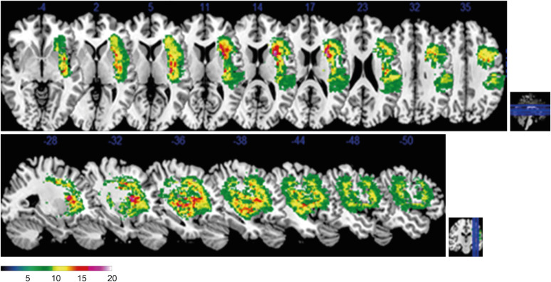
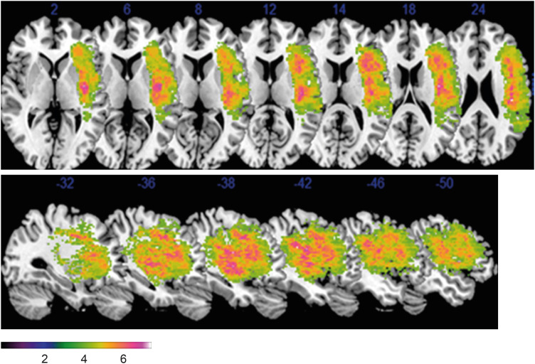
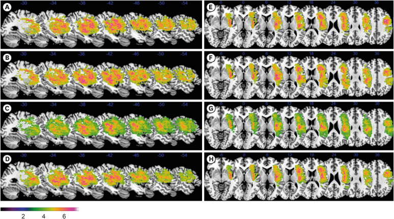
It is unclear how these brain lesions fit into the language processing in acute stroke. In this study, we aimed to investigate the neuroanatomical lesion related to language processing in acute stage of stroke patients using voxel-based lesion-symptom mapping (VLSM). 73 acute first-ever post-stroke patients were enrolled in this retrospective study, who had undertaken brain magnetic resonance imaging (MRI) and Korean version of the Western Aphasia Test within 1 month from onset. Each voxel was compared with aphasia quotient and subtest scores as dependent variables using VLSM. The aphasia group showed significantly much more involvement of extra-nuclear area, insula, inferior frontal gyrus and superior temporal gyrus compared to non-aphasia group. The deficit of spontaneous speech domain was associated with the inferior parietal lobule, inferior and middle frontal gyrus and insula. The insular cortex, inferior parietal lobule, inferior frontal gyrus, middle frontal gyrus and superior temporal gyrus were related to deficit of comprehension. The inferior parietal lobule, insula, precentral gyrus, inferior frontal gyrus were related to the deficit of repetition. The deficit of naming was related to inferior parietal lobule, insula and inferior frontal gyrus. In conclusion, VLSM from early MRI imaging study after stroke may be useful to understand the language process network and establish early rehabilitation strategies after stroke.

 求助内容:
求助内容: 应助结果提醒方式:


