Alexander N. Perez , Kayvon F. Sharif , Erica Guelfi , Sophie Li , Alexis Miller , Kavita Prasad , Robert J. Sinard , James S. Lewis Jr , Michael C. Topf
{"title":"Ex vivo 3D scanning and specimen mapping in anatomic pathology","authors":"Alexander N. Perez , Kayvon F. Sharif , Erica Guelfi , Sophie Li , Alexis Miller , Kavita Prasad , Robert J. Sinard , James S. Lewis Jr , Michael C. Topf","doi":"10.1016/j.jpi.2022.100186","DOIUrl":null,"url":null,"abstract":"<div><p>Structured light three-dimensional (3D) scanning is a ubiquitous mainstay of object inspection and quality control in industrial manufacturing, and has recently been integrated into various medical disciplines. Photorealistic 3D scans can readily be acquired from fresh or formalin-fixed tissue and have potential for use within anatomic pathology (AP) in a variety of scenarios, ranging from direct clinical care to documentation and education. Methods for scanning and post-processing of fresh surgical specimens rely on relatively low-cost and technically simple procedures. Here, we demonstrate potential use of 3D scanning in surgical pathology in the form of a mixed media pathology report with a novel post-scan virtual inking and marking technique to precisely demarcate areas of tissue sectioning and details of final tumor and margin status. We display a sample mixed-media pathology report (3D specimen map) which integrates 3D and conventional pathology reporting methods. Finally, we describe the potential utility of 3D specimen modeling in both didactic and experiential teaching of gross pathology lab procedures.</p></div>","PeriodicalId":37769,"journal":{"name":"Journal of Pathology Informatics","volume":"14 ","pages":"Article 100186"},"PeriodicalIF":0.0000,"publicationDate":"2023-01-01","publicationTypes":"Journal Article","fieldsOfStudy":null,"isOpenAccess":false,"openAccessPdf":"https://www.ncbi.nlm.nih.gov/pmc/articles/PMC9852486/pdf/","citationCount":"5","resultStr":null,"platform":"Semanticscholar","paperid":null,"PeriodicalName":"Journal of Pathology Informatics","FirstCategoryId":"1085","ListUrlMain":"https://www.sciencedirect.com/science/article/pii/S2153353922007866","RegionNum":0,"RegionCategory":null,"ArticlePicture":[],"TitleCN":null,"AbstractTextCN":null,"PMCID":null,"EPubDate":"","PubModel":"","JCR":"Q2","JCRName":"Medicine","Score":null,"Total":0}
引用次数: 5
Abstract
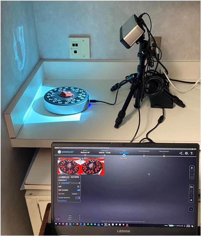
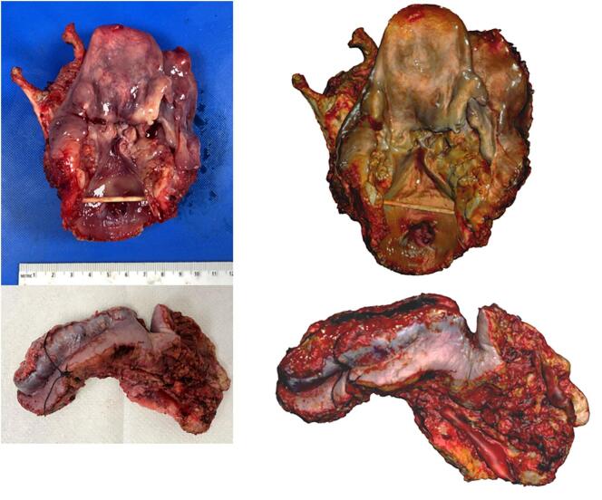
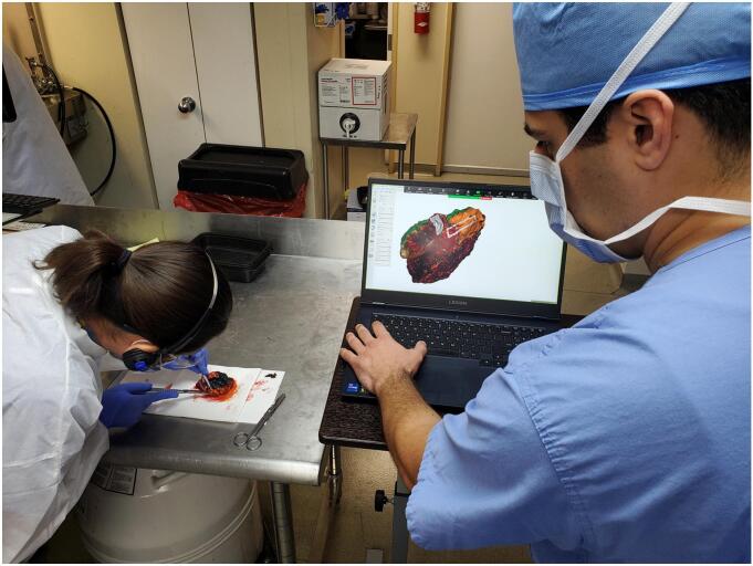
Structured light three-dimensional (3D) scanning is a ubiquitous mainstay of object inspection and quality control in industrial manufacturing, and has recently been integrated into various medical disciplines. Photorealistic 3D scans can readily be acquired from fresh or formalin-fixed tissue and have potential for use within anatomic pathology (AP) in a variety of scenarios, ranging from direct clinical care to documentation and education. Methods for scanning and post-processing of fresh surgical specimens rely on relatively low-cost and technically simple procedures. Here, we demonstrate potential use of 3D scanning in surgical pathology in the form of a mixed media pathology report with a novel post-scan virtual inking and marking technique to precisely demarcate areas of tissue sectioning and details of final tumor and margin status. We display a sample mixed-media pathology report (3D specimen map) which integrates 3D and conventional pathology reporting methods. Finally, we describe the potential utility of 3D specimen modeling in both didactic and experiential teaching of gross pathology lab procedures.
期刊介绍:
The Journal of Pathology Informatics (JPI) is an open access peer-reviewed journal dedicated to the advancement of pathology informatics. This is the official journal of the Association for Pathology Informatics (API). The journal aims to publish broadly about pathology informatics and freely disseminate all articles worldwide. This journal is of interest to pathologists, informaticians, academics, researchers, health IT specialists, information officers, IT staff, vendors, and anyone with an interest in informatics. We encourage submissions from anyone with an interest in the field of pathology informatics. We publish all types of papers related to pathology informatics including original research articles, technical notes, reviews, viewpoints, commentaries, editorials, symposia, meeting abstracts, book reviews, and correspondence to the editors. All submissions are subject to rigorous peer review by the well-regarded editorial board and by expert referees in appropriate specialties.

 求助内容:
求助内容: 应助结果提醒方式:


