Losartan Reduces Remodeling and Apoptosis in an Adriamycin-Induced Cardiomyopathy Rat Model.

Q4 Medicine
Journal of Chest Surgery Pub Date : 2023-09-05 Epub Date: 2023-08-14 DOI:10.5090/jcs.23.044
Hyeon A Kim, Kwan Chang Kim, Hyeryon Lee, Young Mi Hong
{"title":"Losartan Reduces Remodeling and Apoptosis in an Adriamycin-Induced Cardiomyopathy Rat Model.","authors":"Hyeon A Kim, Kwan Chang Kim, Hyeryon Lee, Young Mi Hong","doi":"10.5090/jcs.23.044","DOIUrl":null,"url":null,"abstract":"Background The use of Adriamycin (ADR), also known as doxorubicin, as a chemotherapy agent is limited by its detrimental adverse effects, especially cardiotoxicity. Recent studies have emphasized the crucial role of angiotensin II (Ang-II) in the development of ADR-induced cardiomyopathy. This study aimed to explore the potential cardioprotective effects of losartan in a rat model of ADR-induced cardiomyopathy. Methods Male Sprague-Dawley rats were randomly divided into 3 groups a control group (group C), an ADR-treated group (ADR 5 mg/kg/wk for 3 weeks via intraperitoneal injections; group A), and co-treatment of ADR with losartan group (same dose of ADR and losartan; 10 mg/kg/day per oral for 3 weeks; group L). Western blot analysis was conducted to demonstrate changes in brain natriuretic peptide, collagen 1, tumor necrosis factor (TNF)-α, interleukin-6, matrix metalloproteinase (MMP)-2, B-cell leukemia/lymphoma (Bcl)-2, Bcl-2-associated X (Bax), and caspase-3 protein expression levels in left ventricular (LV) tissues from each group. Results Losartan administration reduced LV hypertrophy, collagen content, and the expression of pro-inflammatory factors TNF-α and MMP-2 in LV tissue. In addition, losartan led to a decrease in the expression of the pro-apoptotic proteins Bax and caspase-3 and an increase in the expression of the anti-apoptotic protein Bcl-2. Moreover, losartan treatment induced a reduction in the apoptotic area compared to group A. Conclusion In an ADR-induced cardiomyopathy rat model, co-administration of ADR with losartan presented cardioprotective effects by attenuating LV hypertrophy, pro-inflammatory factors, and apoptosis in LV tissue.","PeriodicalId":34499,"journal":{"name":"Journal of Chest Surgery","volume":null,"pages":null},"PeriodicalIF":0.0000,"publicationDate":"2023-09-05","publicationTypes":"Journal Article","fieldsOfStudy":null,"isOpenAccess":false,"openAccessPdf":"https://ftp.ncbi.nlm.nih.gov/pub/pmc/oa_pdf/f0/80/jcs-56-5-295.PMC10480395.pdf","citationCount":"0","resultStr":null,"platform":"Semanticscholar","paperid":null,"PeriodicalName":"Journal of Chest Surgery","FirstCategoryId":"1085","ListUrlMain":"https://doi.org/10.5090/jcs.23.044","RegionNum":0,"RegionCategory":null,"ArticlePicture":[],"TitleCN":null,"AbstractTextCN":null,"PMCID":null,"EPubDate":"2023/8/14 0:00:00","PubModel":"Epub","JCR":"Q4","JCRName":"Medicine","Score":null,"Total":0}
引用次数: 0

Abstract

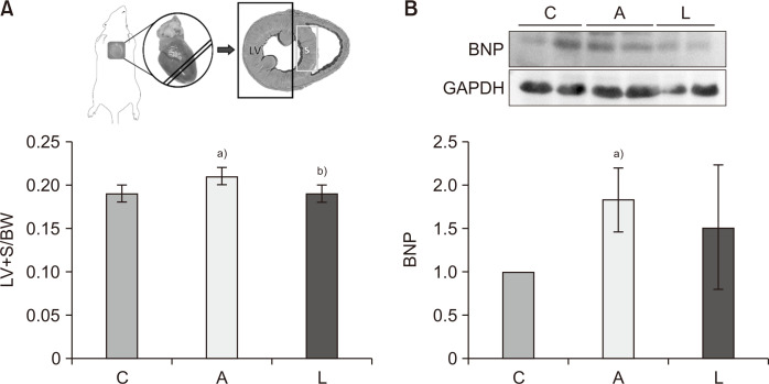
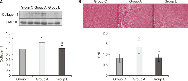
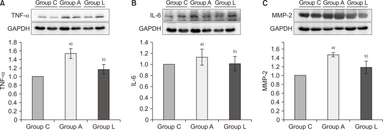
Background The use of Adriamycin (ADR), also known as doxorubicin, as a chemotherapy agent is limited by its detrimental adverse effects, especially cardiotoxicity. Recent studies have emphasized the crucial role of angiotensin II (Ang-II) in the development of ADR-induced cardiomyopathy. This study aimed to explore the potential cardioprotective effects of losartan in a rat model of ADR-induced cardiomyopathy. Methods Male Sprague-Dawley rats were randomly divided into 3 groups a control group (group C), an ADR-treated group (ADR 5 mg/kg/wk for 3 weeks via intraperitoneal injections; group A), and co-treatment of ADR with losartan group (same dose of ADR and losartan; 10 mg/kg/day per oral for 3 weeks; group L). Western blot analysis was conducted to demonstrate changes in brain natriuretic peptide, collagen 1, tumor necrosis factor (TNF)-α, interleukin-6, matrix metalloproteinase (MMP)-2, B-cell leukemia/lymphoma (Bcl)-2, Bcl-2-associated X (Bax), and caspase-3 protein expression levels in left ventricular (LV) tissues from each group. Results Losartan administration reduced LV hypertrophy, collagen content, and the expression of pro-inflammatory factors TNF-α and MMP-2 in LV tissue. In addition, losartan led to a decrease in the expression of the pro-apoptotic proteins Bax and caspase-3 and an increase in the expression of the anti-apoptotic protein Bcl-2. Moreover, losartan treatment induced a reduction in the apoptotic area compared to group A. Conclusion In an ADR-induced cardiomyopathy rat model, co-administration of ADR with losartan presented cardioprotective effects by attenuating LV hypertrophy, pro-inflammatory factors, and apoptosis in LV tissue.
Abstract Image

Abstract Image

Abstract Image

氯沙坦减少阿霉素诱导的心肌病大鼠模型的重塑和细胞凋亡。
背景:阿霉素(ADR),又称阿霉素,作为一种化疗药物,由于其有害的副作用,尤其是心脏毒性而受到限制。最近的研究强调了血管紧张素II (Ang-II)在adr诱导的心肌病发展中的关键作用。本研究旨在探讨氯沙坦对不良反应性心肌病大鼠模型的潜在心脏保护作用。方法:雄性Sprague-Dawley大鼠随机分为3组:对照组(C组)、ADR处理组(ADR 5 mg/kg/周,腹腔注射3周;A组),ADR与氯沙坦联合治疗组(ADR与氯沙坦剂量相同;10mg /kg/天,口服3周;Western blot分析各组左心室(LV)组织中脑利钠肽、胶原蛋白1、肿瘤坏死因子(TNF)-α、白细胞介素-6、基质金属蛋白酶(MMP)-2、b细胞白血病/淋巴瘤(Bcl)-2、Bcl-2相关X (Bax)和caspase-3蛋白表达水平的变化。结果:氯沙坦可降低左室肥大、胶原含量及促炎因子TNF-α、MMP-2在左室组织中的表达。此外,氯沙坦导致促凋亡蛋白Bax和caspase-3的表达减少,抗凋亡蛋白Bcl-2的表达增加。此外,与a组相比,氯沙坦治疗导致凋亡面积减少。结论:在ADR诱导的心肌病大鼠模型中,ADR与氯沙坦共给药通过减轻左室肥厚、促炎因子和左室组织凋亡而具有心脏保护作用。
本文章由计算机程序翻译,如有差异,请以英文原文为准。
求助全文
约1分钟内获得全文 求助全文
来源期刊
Journal of Chest Surgery
Journal of Chest Surgery Medicine-Surgery
CiteScore
0.80
自引率
0.00%
发文量
76
审稿时长
7 weeks
×
引用
GB/T 7714-2015
复制
MLA
复制
APA
复制
导出至
BibTeX EndNote RefMan NoteFirst NoteExpress
×
提示
您的信息不完整,为了账户安全,请先补充。
现在去补充
×
提示
您因"违规操作"
具体请查看互助需知
我知道了
×
提示
确定
请完成安全验证×
copy
已复制链接
快去分享给好友吧!
我知道了
右上角分享
点击右上角分享
0
联系我们:info@booksci.cn Book学术提供免费学术资源搜索服务,方便国内外学者检索中英文文献。致力于提供最便捷和优质的服务体验。 Copyright © 2023 布克学术 All rights reserved.
京ICP备2023020795号-1
ghs 京公网安备 11010802042870号
Book学术文献互助
Book学术文献互助群
群 号:481959085
Book学术官方微信