Aladár David Rónaszéki, Ibolyka Dudás, Boglarka Zsély, Bettina Katalin Budai, Róbert Stollmayer, Oszkár Hahn, Barbara Csongrády, Byung-So Park, Pál Maurovich-Horvat, Gabriella Győri, Pal Novak Kaposi
{"title":"Microvascular flow imaging to differentiate focal hepatic lesions: the spoke-wheel pattern as a specific sign of focal nodular hyperplasia.","authors":"Aladár David Rónaszéki, Ibolyka Dudás, Boglarka Zsély, Bettina Katalin Budai, Róbert Stollmayer, Oszkár Hahn, Barbara Csongrády, Byung-So Park, Pál Maurovich-Horvat, Gabriella Győri, Pal Novak Kaposi","doi":"10.14366/usg.22028","DOIUrl":null,"url":null,"abstract":"<p><p>Microvascular flow imaging (MVFI) is an advanced Doppler ultrasound technique designed to detect slow-velocity blood flow in small-caliber microvessels. This technique is capable of realtime, highly detailed visualization of tumor vessels without using a contrast agent. MVFI has been recently applied for the characterization of focal liver lesions and has revealed typical vascularity distributions in multiple types thereof. Focal nodular hyperplasia (FNH) constitutes an important differential diagnosis of malignant liver tumors. In this essay, we provide iconographic documentation of the MVFI appearance of FNH and other common solid liver lesions. Identifying the typical patterns of vascularity, including the spoke-wheel pattern with MVFI, can expedite the diagnosis, spare patients from unnecessary procedures, and save costs.</p>","PeriodicalId":54227,"journal":{"name":"Ultrasonography","volume":"42 1","pages":"172-181"},"PeriodicalIF":2.4000,"publicationDate":"2023-01-01","publicationTypes":"Journal Article","fieldsOfStudy":null,"isOpenAccess":false,"openAccessPdf":"https://ftp.ncbi.nlm.nih.gov/pub/pmc/oa_pdf/0f/ba/usg-22028.PMC9816699.pdf","citationCount":"2","resultStr":null,"platform":"Semanticscholar","paperid":null,"PeriodicalName":"Ultrasonography","FirstCategoryId":"3","ListUrlMain":"https://doi.org/10.14366/usg.22028","RegionNum":3,"RegionCategory":"医学","ArticlePicture":[],"TitleCN":null,"AbstractTextCN":null,"PMCID":null,"EPubDate":"","PubModel":"","JCR":"Q2","JCRName":"RADIOLOGY, NUCLEAR MEDICINE & MEDICAL IMAGING","Score":null,"Total":0}
引用次数: 2
Abstract
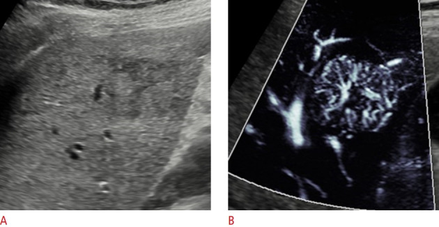
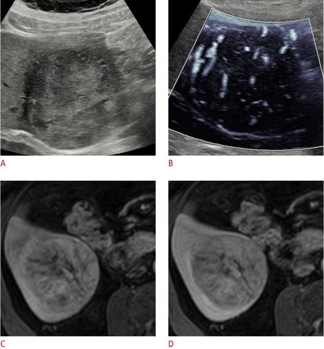
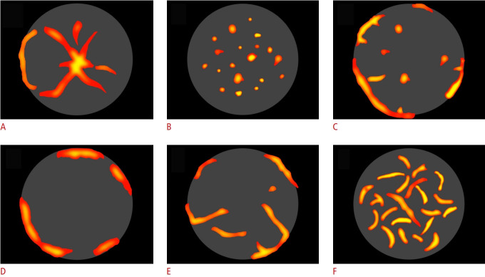
Microvascular flow imaging (MVFI) is an advanced Doppler ultrasound technique designed to detect slow-velocity blood flow in small-caliber microvessels. This technique is capable of realtime, highly detailed visualization of tumor vessels without using a contrast agent. MVFI has been recently applied for the characterization of focal liver lesions and has revealed typical vascularity distributions in multiple types thereof. Focal nodular hyperplasia (FNH) constitutes an important differential diagnosis of malignant liver tumors. In this essay, we provide iconographic documentation of the MVFI appearance of FNH and other common solid liver lesions. Identifying the typical patterns of vascularity, including the spoke-wheel pattern with MVFI, can expedite the diagnosis, spare patients from unnecessary procedures, and save costs.
UltrasonographyMedicine-Radiology, Nuclear Medicine and Imaging
CiteScore
5.10
自引率
6.50%
发文量
78
审稿时长
15 weeks
期刊介绍:
Ultrasonography, the official English-language journal of the Korean Society of Ultrasound in Medicine (KSUM), is an international peer-reviewed academic journal dedicated to practice, research, technology, and education dealing with medical ultrasound. It is renamed from the Journal of Korean Society of Ultrasound in Medicine in January 2014, and published four times per year: January 1, April 1, July 1, and October 1. Original articles, technical notes, topical reviews, perspectives, pictorial essays, and timely editorial materials are published in Ultrasonography covering state-of-the-art content.
Ultrasonography aims to provide updated information on new diagnostic concepts and technical developments, including experimental animal studies using new equipment in addition to well-designed reviews of contemporary issues in patient care. Along with running KSUM Open, the annual international congress of KSUM, Ultrasonography also serves as a medium for cooperation among physicians and specialists from around the world who are focusing on various ultrasound technology and disease problems and relevant basic science.
 求助内容:
求助内容: 应助结果提醒方式:


