{"title":"Evaluation of the different exposure parameters for the accurate diagnosis of peri-implantitis severity in digital panoramic radiography.","authors":"E Sadik, C Gökmenoğlu, G Altun, C Kara","doi":"10.4317/medoral.25501","DOIUrl":null,"url":null,"abstract":"<p><strong>Background: </strong>To evaluate the accuracy of the diagnosis of peri-implant bone defects' severities in digital panoramic radiographs obtained at different tube voltage and/or tube current settings.</p><p><strong>Material and methods: </strong>Two different sizes of peri-implant bone defects (type 1 and type 2) were prepared after the implants were inserted into 29 bovine rib blocks. Digital panoramic radiographs were obtained at eight different tube voltage and/or tube current settings for all rib blocks. Implant images were cropped separately. The average intensity value (AIV) of cropped images were analyzed using Adobe Photoshop CC software. The Kruskal-Wallis H test was used to compare AIVs. All cropped images were evaluated using a five-point Likert scale for the likelihood of a bone defect being absent or present. The weighted kappa values were calculated to compare observer agreement and ROC analysis was performed to determine the appropriate exposure parameters.</p><p><strong>Results: </strong>The lowest AIV was obtained at 72 kV/6.3 mA (92.162±16.016), and the highest AIV was obtained at 60 kV/3.2 mA (179.050±13.823). The Kruskal-Wallis H test showed significant differences in the AIVs according to the exposure parameters (p<0.001). The kappa coefficient for the inter-observer agreement was excellent (0.864, p<0.001). The AUC values for type 1 defects ranged from 0.778 and 0.860; for type 2 defects ranged from 0.920 and 0.967. The AUC value of type 1 defects was slightly better in panoramic images obtained with high kV and low mA levels (72 kV/3.2 mA), compared to others.</p><p><strong>Conclusions: </strong>In daily clinical routine, peri-implant bone defects might be evaluated by panoramic radiographs obtained with all kV and mA values tested. However, to avoid misdiagnosing and for better accuracy, panoramic radiographs obtained with high kV and low mA levels suitable for patients should be used, especially in the detection of small or initial bone defects.</p>","PeriodicalId":18351,"journal":{"name":"Medicina oral, patologia oral y cirugia bucal","volume":"28 1","pages":"e16-e24"},"PeriodicalIF":2.1000,"publicationDate":"2023-01-01","publicationTypes":"Journal Article","fieldsOfStudy":null,"isOpenAccess":false,"openAccessPdf":"https://www.ncbi.nlm.nih.gov/pmc/articles/PMC9805328/pdf/","citationCount":"0","resultStr":null,"platform":"Semanticscholar","paperid":null,"PeriodicalName":"Medicina oral, patologia oral y cirugia bucal","FirstCategoryId":"3","ListUrlMain":"https://doi.org/10.4317/medoral.25501","RegionNum":3,"RegionCategory":"医学","ArticlePicture":[],"TitleCN":null,"AbstractTextCN":null,"PMCID":null,"EPubDate":"","PubModel":"","JCR":"","JCRName":"","Score":null,"Total":0}
引用次数: 0
Abstract
Background: To evaluate the accuracy of the diagnosis of peri-implant bone defects' severities in digital panoramic radiographs obtained at different tube voltage and/or tube current settings.
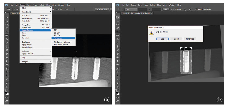
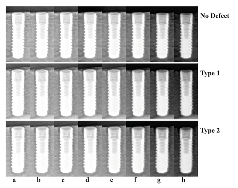
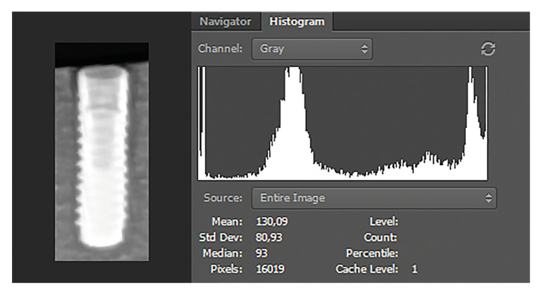
Material and methods: Two different sizes of peri-implant bone defects (type 1 and type 2) were prepared after the implants were inserted into 29 bovine rib blocks. Digital panoramic radiographs were obtained at eight different tube voltage and/or tube current settings for all rib blocks. Implant images were cropped separately. The average intensity value (AIV) of cropped images were analyzed using Adobe Photoshop CC software. The Kruskal-Wallis H test was used to compare AIVs. All cropped images were evaluated using a five-point Likert scale for the likelihood of a bone defect being absent or present. The weighted kappa values were calculated to compare observer agreement and ROC analysis was performed to determine the appropriate exposure parameters.
Results: The lowest AIV was obtained at 72 kV/6.3 mA (92.162±16.016), and the highest AIV was obtained at 60 kV/3.2 mA (179.050±13.823). The Kruskal-Wallis H test showed significant differences in the AIVs according to the exposure parameters (p<0.001). The kappa coefficient for the inter-observer agreement was excellent (0.864, p<0.001). The AUC values for type 1 defects ranged from 0.778 and 0.860; for type 2 defects ranged from 0.920 and 0.967. The AUC value of type 1 defects was slightly better in panoramic images obtained with high kV and low mA levels (72 kV/3.2 mA), compared to others.
Conclusions: In daily clinical routine, peri-implant bone defects might be evaluated by panoramic radiographs obtained with all kV and mA values tested. However, to avoid misdiagnosing and for better accuracy, panoramic radiographs obtained with high kV and low mA levels suitable for patients should be used, especially in the detection of small or initial bone defects.
期刊介绍:
1. Oral Medicine and Pathology:
Clinicopathological as well as medical or surgical management aspects of
diseases affecting oral mucosa, salivary glands, maxillary bones, as well as
orofacial neurological disorders, and systemic conditions with an impact on
the oral cavity.
2. Oral Surgery:
Surgical management aspects of diseases affecting oral mucosa, salivary glands,
maxillary bones, teeth, implants, oral surgical procedures. Surgical management
of diseases affecting head and neck areas.
3. Medically compromised patients in Dentistry:
Articles discussing medical problems in Odontology will also be included, with
a special focus on the clinico-odontological management of medically compromised patients, and considerations regarding high-risk or disabled patients.
4. Implantology
5. Periodontology
 求助内容:
求助内容: 应助结果提醒方式:


