K Gobi, Venkatesh Kasi Arunachalam, Rajesh Kumar Varatharajaperumal, Mathew Cherian, Gopinath Periaswamy, S Rajesh
{"title":"The role of ultra-low-dose computed tomography in the detection of pulmonary pathologies: a prospective observational study.","authors":"K Gobi, Venkatesh Kasi Arunachalam, Rajesh Kumar Varatharajaperumal, Mathew Cherian, Gopinath Periaswamy, S Rajesh","doi":"10.5114/pjr.2022.121433","DOIUrl":null,"url":null,"abstract":"<p><strong>Purpose: </strong>The aim of the study was to compare the image noise, radiation dose, and image quality of ultra-low-dose computed tomography (CT) and standard CT in the imaging of pulmonary pathologies.</p><p><strong>Material and methods: </strong>This observational study was performed between July 2020 and August 2021. All enrolled patients underwent both ultra-low-dose and standard CTs. The image noise, image quality for normal pulmonary structures, presence or absence of various pulmonary lesions, and radiation dose were recorded for each of the scans. The findings of standard-dose CT were regarded as the gold standard and compared with that of ultra-low-dose CT.</p><p><strong>Results: </strong>A total of 124 patients were included in the study. The image noise was higher in the ultra-low-dose CT compared to standard-dose CT. The overall image quality was determined to be diagnostic in 100% of standard CT images and in 96.77% of ultra-low-dose CT images with proportional worsening of the image quality as the body mass index (BMI) range was increased. Ultra-low-dose CT offered higher (> 90%) sensitivity for lesions like consolidation (97%), pleural effusion (95%), fibrosis (92%), and solid pulmonary nodules (91%). The effective radiation dose (mSv) was many times lower in ultra-low-dose CT when compared to standard-dose CT (mean ± SD: 0.50 ± 0.005 vs. 3.99 ± 1.57).</p><p><strong>Conclusions: </strong>The radiation dose of ultra-low-dose chest CT was almost equal to that of a chest X-ray. It could be used for the screening and/or follow-up of patients with solid pulmonary nodules (> 3 mm) and consolidation.</p>","PeriodicalId":47128,"journal":{"name":"Polish Journal of Radiology","volume":"87 ","pages":"e597-e605"},"PeriodicalIF":0.9000,"publicationDate":"2022-01-01","publicationTypes":"Journal Article","fieldsOfStudy":null,"isOpenAccess":false,"openAccessPdf":"https://ftp.ncbi.nlm.nih.gov/pub/pmc/oa_pdf/74/7c/PJR-87-48263.PMC9749781.pdf","citationCount":"0","resultStr":null,"platform":"Semanticscholar","paperid":null,"PeriodicalName":"Polish Journal of Radiology","FirstCategoryId":"1085","ListUrlMain":"https://doi.org/10.5114/pjr.2022.121433","RegionNum":0,"RegionCategory":null,"ArticlePicture":[],"TitleCN":null,"AbstractTextCN":null,"PMCID":null,"EPubDate":"","PubModel":"","JCR":"Q4","JCRName":"RADIOLOGY, NUCLEAR MEDICINE & MEDICAL IMAGING","Score":null,"Total":0}
引用次数: 0
Abstract
Purpose: The aim of the study was to compare the image noise, radiation dose, and image quality of ultra-low-dose computed tomography (CT) and standard CT in the imaging of pulmonary pathologies.
Material and methods: This observational study was performed between July 2020 and August 2021. All enrolled patients underwent both ultra-low-dose and standard CTs. The image noise, image quality for normal pulmonary structures, presence or absence of various pulmonary lesions, and radiation dose were recorded for each of the scans. The findings of standard-dose CT were regarded as the gold standard and compared with that of ultra-low-dose CT.
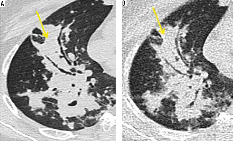
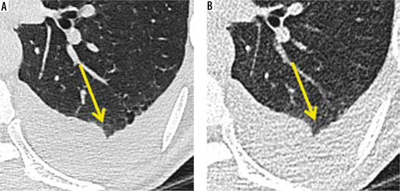
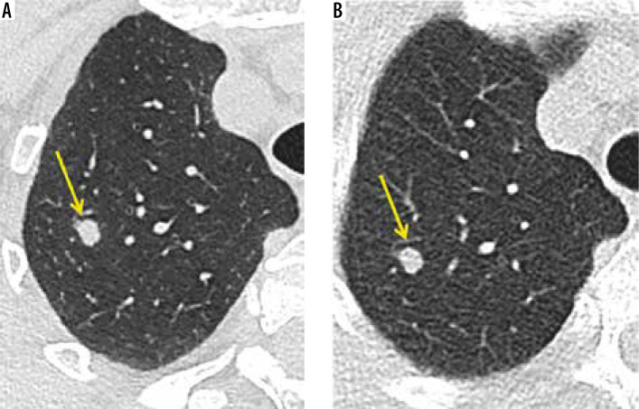
Results: A total of 124 patients were included in the study. The image noise was higher in the ultra-low-dose CT compared to standard-dose CT. The overall image quality was determined to be diagnostic in 100% of standard CT images and in 96.77% of ultra-low-dose CT images with proportional worsening of the image quality as the body mass index (BMI) range was increased. Ultra-low-dose CT offered higher (> 90%) sensitivity for lesions like consolidation (97%), pleural effusion (95%), fibrosis (92%), and solid pulmonary nodules (91%). The effective radiation dose (mSv) was many times lower in ultra-low-dose CT when compared to standard-dose CT (mean ± SD: 0.50 ± 0.005 vs. 3.99 ± 1.57).
Conclusions: The radiation dose of ultra-low-dose chest CT was almost equal to that of a chest X-ray. It could be used for the screening and/or follow-up of patients with solid pulmonary nodules (> 3 mm) and consolidation.

 求助内容:
求助内容: 应助结果提醒方式:


