Wilson Sharp, Isabel Jang, Michael J Diaz, Leila C Tou, Charles A Agyemang, Rebekah E Carter, Sarah Sevigny, Muhammad Umair
{"title":"Comparative imaging findings among different primary betacoronaviruses.","authors":"Wilson Sharp, Isabel Jang, Michael J Diaz, Leila C Tou, Charles A Agyemang, Rebekah E Carter, Sarah Sevigny, Muhammad Umair","doi":"10.5114/pjr.2022.121763","DOIUrl":null,"url":null,"abstract":"<p><strong>Purpose: </strong>Coronaviruses (CoV) are single-stranded RNA viruses that transmit from animal species to humans, causing a threat to global health. We aim to summarize common imaging findings of 3 betacoronaviruses (b-CoVs) and the common clinical manifestation, to provide a better understanding of the courses of the disease.</p><p><strong>Material and methods: </strong>The Pubmed and Google Scholar databases were searched for the terms \"SARS-CoV\" OR \"COVID-19\" OR \"MERS-CoV\". Imaging-specific searches included keyword searches for \"CT\" AND \"imaging\". Clinical presentation-specific searches included keyword searches for \"clinical\" AND \"manifestation\" AND \"cardio-vascular\" OR \"neurology\" OR \"gastrointestinal\" OR \"hematology\". In total, 77 articles were selected for discussion in the current literature review.</p><p><strong>Results: </strong>Human b-CoVs infection presented consistent indications of ground-glass opacities (GGO), consolidation, and interlobular septal thickening. Pleural effusion was also common in all 3 b-CoVs, but it was least present in SARS-CoV-2 infection. Bilateral lung involvement was common to both MERS-CoV and SARS-CoV-2 infection. Cardiovascular, neurological, haematological, and gastrointestinal were common clinical presentations found in patients infected with b-CoVs.</p><p><strong>Conclusions: </strong>The comparison of imaging findings can be applied in clinical practice to distinguish the 3 CoV through different imaging modalities. It is crucial to understand the possible imaging findings and clinical presentations to better understand the course of the disease as well as prepare for future variants.</p>","PeriodicalId":47128,"journal":{"name":"Polish Journal of Radiology","volume":"87 ","pages":"e641-e651"},"PeriodicalIF":0.9000,"publicationDate":"2022-01-01","publicationTypes":"Journal Article","fieldsOfStudy":null,"isOpenAccess":false,"openAccessPdf":"https://ftp.ncbi.nlm.nih.gov/pub/pmc/oa_pdf/b0/fb/PJR-87-48383.PMC9749786.pdf","citationCount":"0","resultStr":null,"platform":"Semanticscholar","paperid":null,"PeriodicalName":"Polish Journal of Radiology","FirstCategoryId":"1085","ListUrlMain":"https://doi.org/10.5114/pjr.2022.121763","RegionNum":0,"RegionCategory":null,"ArticlePicture":[],"TitleCN":null,"AbstractTextCN":null,"PMCID":null,"EPubDate":"","PubModel":"","JCR":"Q4","JCRName":"RADIOLOGY, NUCLEAR MEDICINE & MEDICAL IMAGING","Score":null,"Total":0}
引用次数: 0
Abstract
Purpose: Coronaviruses (CoV) are single-stranded RNA viruses that transmit from animal species to humans, causing a threat to global health. We aim to summarize common imaging findings of 3 betacoronaviruses (b-CoVs) and the common clinical manifestation, to provide a better understanding of the courses of the disease.
Material and methods: The Pubmed and Google Scholar databases were searched for the terms "SARS-CoV" OR "COVID-19" OR "MERS-CoV". Imaging-specific searches included keyword searches for "CT" AND "imaging". Clinical presentation-specific searches included keyword searches for "clinical" AND "manifestation" AND "cardio-vascular" OR "neurology" OR "gastrointestinal" OR "hematology". In total, 77 articles were selected for discussion in the current literature review.
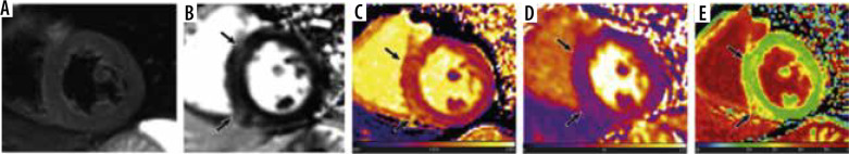
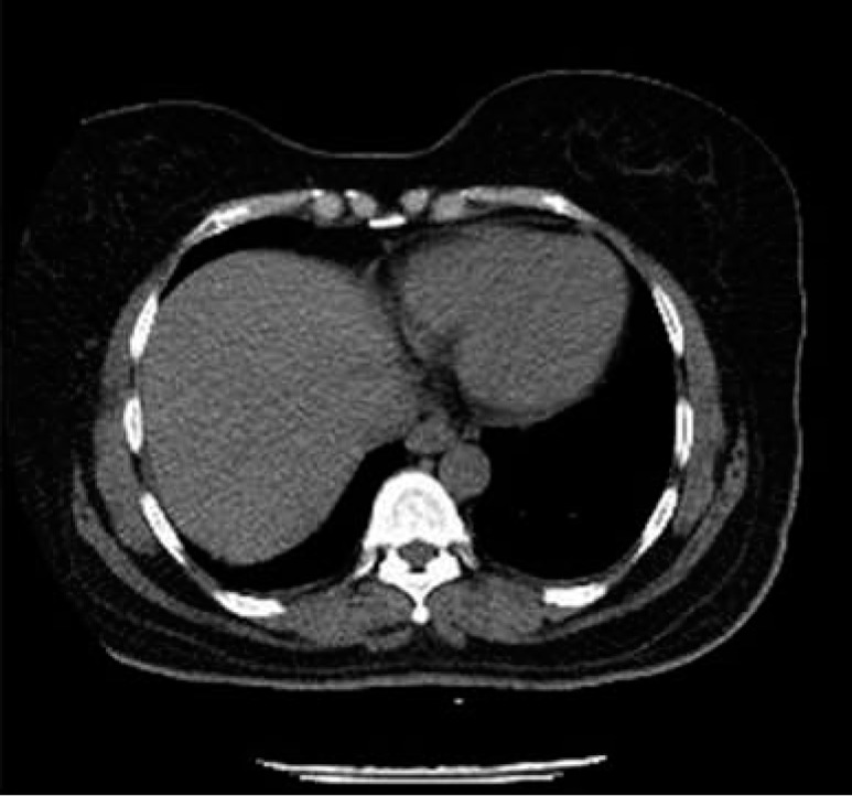
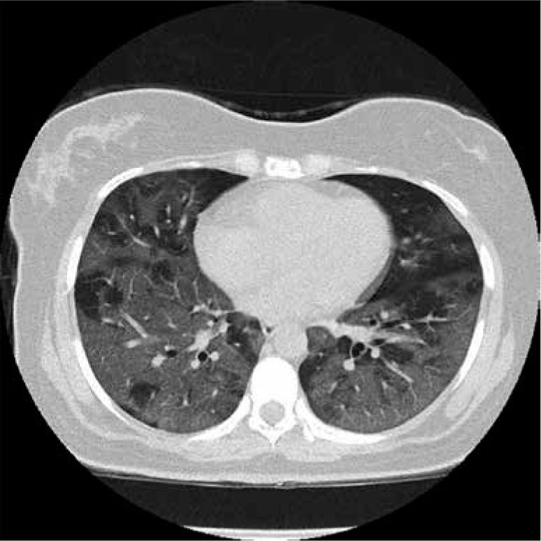
Results: Human b-CoVs infection presented consistent indications of ground-glass opacities (GGO), consolidation, and interlobular septal thickening. Pleural effusion was also common in all 3 b-CoVs, but it was least present in SARS-CoV-2 infection. Bilateral lung involvement was common to both MERS-CoV and SARS-CoV-2 infection. Cardiovascular, neurological, haematological, and gastrointestinal were common clinical presentations found in patients infected with b-CoVs.
Conclusions: The comparison of imaging findings can be applied in clinical practice to distinguish the 3 CoV through different imaging modalities. It is crucial to understand the possible imaging findings and clinical presentations to better understand the course of the disease as well as prepare for future variants.

 求助内容:
求助内容: 应助结果提醒方式:


