Visualizing and understanding inherent features in SD-OCT for the progression of age-related macular degeneration using deconvolutional neural networks
Sajib Saha, Ziyuan Wang, Srinivas Sadda, Yogesan Kanagasingam, Zhihong Hu
{"title":"Visualizing and understanding inherent features in SD-OCT for the progression of age-related macular degeneration using deconvolutional neural networks","authors":"Sajib Saha, Ziyuan Wang, Srinivas Sadda, Yogesan Kanagasingam, Zhihong Hu","doi":"10.1002/ail2.16","DOIUrl":null,"url":null,"abstract":"<p>To develop a convolutional neural network visualization strategy so that optical coherence tomography (OCT) features contributing to the evolution of age-related macular degeneration (AMD) can be better determined. We have trained a U-Net model to utilize baseline OCT to predict the progression of geographic atrophy (GA), a late stage manifestation of AMD. We have augmented the U-Net architecture by attaching deconvolutional neural networks (deconvnets). Deconvnets produce the reconstructed feature maps and provide an indication regarding the inherent baseline OCT features contributing to GA progression. Experiments were conducted on longitudinal spectral domain (SD)-OCT and fundus autofluorescence images collected from 70 eyes with GA. The intensity of Bruch's membrane-outer choroid (BMChoroid) retinal junction exhibited a relative importance of 24%, in the GA progression. The intensity of the inner retinal pigment epithelium (RPE) and BM junction (InRPEBM) showed a relative importance of 22%. BMChoroid (where the AMD feature/damage of choriocapillaris was included) followed by InRPEBM (where the AMD feature/damage of RPE was included) are the layers which appear to be most relevant in predicting the progression of AMD.</p>","PeriodicalId":72253,"journal":{"name":"Applied AI letters","volume":"1 1","pages":""},"PeriodicalIF":0.0000,"publicationDate":"2020-10-14","publicationTypes":"Journal Article","fieldsOfStudy":null,"isOpenAccess":false,"openAccessPdf":"https://sci-hub-pdf.com/10.1002/ail2.16","citationCount":"6","resultStr":null,"platform":"Semanticscholar","paperid":null,"PeriodicalName":"Applied AI letters","FirstCategoryId":"1085","ListUrlMain":"https://onlinelibrary.wiley.com/doi/10.1002/ail2.16","RegionNum":0,"RegionCategory":null,"ArticlePicture":[],"TitleCN":null,"AbstractTextCN":null,"PMCID":null,"EPubDate":"","PubModel":"","JCR":"","JCRName":"","Score":null,"Total":0}
引用次数: 6
Abstract
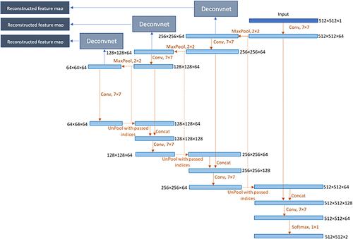
To develop a convolutional neural network visualization strategy so that optical coherence tomography (OCT) features contributing to the evolution of age-related macular degeneration (AMD) can be better determined. We have trained a U-Net model to utilize baseline OCT to predict the progression of geographic atrophy (GA), a late stage manifestation of AMD. We have augmented the U-Net architecture by attaching deconvolutional neural networks (deconvnets). Deconvnets produce the reconstructed feature maps and provide an indication regarding the inherent baseline OCT features contributing to GA progression. Experiments were conducted on longitudinal spectral domain (SD)-OCT and fundus autofluorescence images collected from 70 eyes with GA. The intensity of Bruch's membrane-outer choroid (BMChoroid) retinal junction exhibited a relative importance of 24%, in the GA progression. The intensity of the inner retinal pigment epithelium (RPE) and BM junction (InRPEBM) showed a relative importance of 22%. BMChoroid (where the AMD feature/damage of choriocapillaris was included) followed by InRPEBM (where the AMD feature/damage of RPE was included) are the layers which appear to be most relevant in predicting the progression of AMD.

 求助内容:
求助内容: 应助结果提醒方式:


