Ricardo Ysaac García Núñez, Katherine Rasia Gonzales Córdova, Yuri Karaccas de Carvalho
{"title":"Tridimensional models and radiographic study of dorsal laminectomy and thoracolumbar hemilaminectomy in dogs.","authors":"Ricardo Ysaac García Núñez, Katherine Rasia Gonzales Córdova, Yuri Karaccas de Carvalho","doi":"10.1590/acb382623","DOIUrl":null,"url":null,"abstract":"<p><strong>Purpose: </strong>To create three-dimensional anatomical models of the thoracic and lumbar portions of the canine spine that reproduce the vertebral surgical approaches of dorsal laminectomy and hemilaminectomy, and to perform the respective radiographic evaluations of each approach.</p><p><strong>Methods: </strong>In a digital archive of the canine spine, digitally replicate the dorsal laminectomy and hemilaminectomy in the thoracic and lumbar portions and, then, make tridimensional prints of the vertebral models and obtain radiographs in three dorsoventral, ventrodorsal and laterolateral projections.</p><p><strong>Results: </strong>The anatomical models of the surgical spinal canal accesses of the thoracic and lumbar portions showed great fidelity to the natural bones. The created accesses have the proper shape, location and size, and their radiographic images showed similar radiodensities.</p><p><strong>Conclusions: </strong>The replicas of the dorsal laminectomy and hemilaminectomy developed in the anatomical models in the thoracic and lumbar portions are able to represent the technical recommendations of the specialized literature, as well as their respective radiographic images, which have certain radiological properties that allow to make a deep radiological study. Therefore, the models are useful for neurosurgical training.</p>","PeriodicalId":6992,"journal":{"name":"Acta cirurgica brasileira","volume":"38 ","pages":"e382623"},"PeriodicalIF":1.3000,"publicationDate":"2023-08-04","publicationTypes":"Journal Article","fieldsOfStudy":null,"isOpenAccess":false,"openAccessPdf":"https://www.ncbi.nlm.nih.gov/pmc/articles/PMC10403244/pdf/","citationCount":"0","resultStr":null,"platform":"Semanticscholar","paperid":null,"PeriodicalName":"Acta cirurgica brasileira","FirstCategoryId":"3","ListUrlMain":"https://doi.org/10.1590/acb382623","RegionNum":4,"RegionCategory":"医学","ArticlePicture":[],"TitleCN":null,"AbstractTextCN":null,"PMCID":null,"EPubDate":"2023/1/1 0:00:00","PubModel":"eCollection","JCR":"Q3","JCRName":"SURGERY","Score":null,"Total":0}
引用次数: 0
Abstract
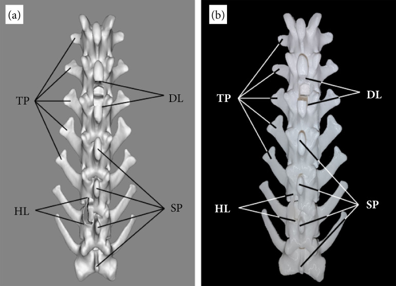
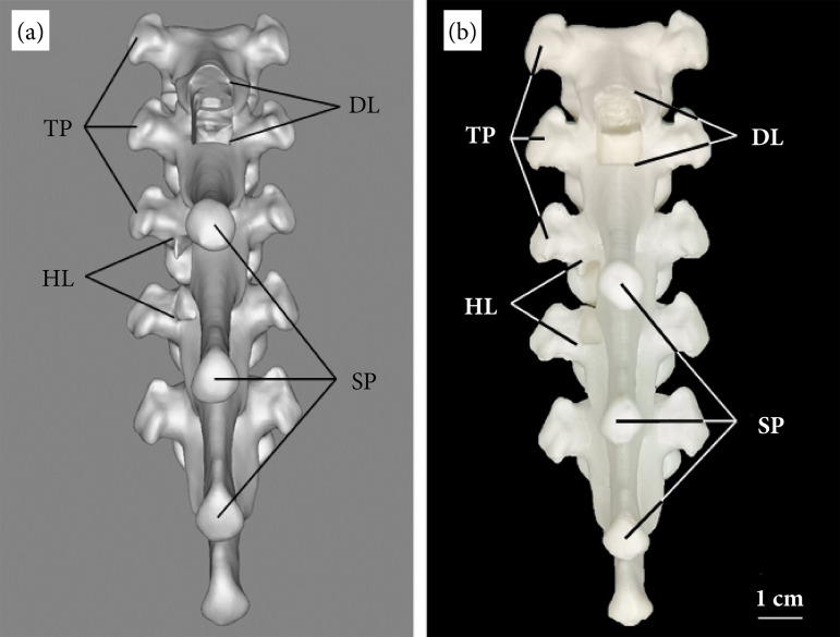
Purpose: To create three-dimensional anatomical models of the thoracic and lumbar portions of the canine spine that reproduce the vertebral surgical approaches of dorsal laminectomy and hemilaminectomy, and to perform the respective radiographic evaluations of each approach.
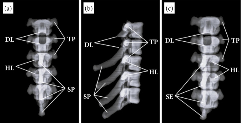
Methods: In a digital archive of the canine spine, digitally replicate the dorsal laminectomy and hemilaminectomy in the thoracic and lumbar portions and, then, make tridimensional prints of the vertebral models and obtain radiographs in three dorsoventral, ventrodorsal and laterolateral projections.
Results: The anatomical models of the surgical spinal canal accesses of the thoracic and lumbar portions showed great fidelity to the natural bones. The created accesses have the proper shape, location and size, and their radiographic images showed similar radiodensities.
Conclusions: The replicas of the dorsal laminectomy and hemilaminectomy developed in the anatomical models in the thoracic and lumbar portions are able to represent the technical recommendations of the specialized literature, as well as their respective radiographic images, which have certain radiological properties that allow to make a deep radiological study. Therefore, the models are useful for neurosurgical training.
 求助内容:
求助内容: 应助结果提醒方式:


