{"title":"Carotid Artery Stenting Using Stent-in-Stent Technique with a Closed-Cell Stent and a Dual-Layer Micromesh Stent: A Case Report.","authors":"Yoshitaka Yamaguchi, Tatsuro Takada, Kazuki Uchida, Kei Miyata, Kota Kurisu, Tomohiro Okuyama, Fumiki Tomeoka, Minoru Ajiki, Masaaki Hokari, Katsuyuki Asaoka","doi":"10.5797/jnet.cr.2023-0003","DOIUrl":null,"url":null,"abstract":"<p><strong>Objective: </strong>Recent studies evaluating plaque protrusion at carotid artery stenting (CAS) using optical coherence tomography showed not a few cases of plaque protrusion when using double-layer micromesh stents. We report a case of symptomatic internal carotid artery (ICA) stenosis with at-risk unstable plaques in which CAS was successfully performed using a stent-in-stent technique by the combined use of a closed-cell stent and a dual-layer micromesh stent.</p><p><strong>Case presentation: </strong>An 87-year-old Japanese man with dysarthria and right hemiparesis was diagnosed with atheromatous cerebral embolism caused by severe left ICA stenosis on MRI and DSA. MRI with T1-weighted black blood methods showed high intensities in the plaques of the left ICA, suggesting unstable plaque characteristics with intraplaque hemorrhage components. On day 20, CAS was performed. After the pre-stent dilation under proximal and distal protection, a Carotid WALLSTENT was placed to cover the stenotic lesion. Then, a CASPER Rx was placed from the proximal left ICA to the common carotid artery to cover the Carotid WALLSTENT. Although visible plaque debris was recognized in the aspirated blood, the debris became invisible after aspiration of 1300 mL. Postoperative angiography showed enough dilation of the left ICA, with no plaque protrusion or acute stent thrombosis. The patient had an uneventful postoperative course and was discharged without any neurological sequelae.</p><p><strong>Conclusion: </strong>The present case suggests that the combined stent-in-stent technique using a closed-cell stent and a micromesh stent can be considered as one of the treatment strategies for preventing plaque protrusion and procedural ischemic complications in patients with high-risk carotid plaques.</p>","PeriodicalId":73856,"journal":{"name":"Journal of neuroendovascular therapy","volume":"17 5","pages":"101-106"},"PeriodicalIF":0.0000,"publicationDate":"2023-01-01","publicationTypes":"Journal Article","fieldsOfStudy":null,"isOpenAccess":false,"openAccessPdf":"https://ftp.ncbi.nlm.nih.gov/pub/pmc/oa_pdf/a2/18/jnet-17-101.PMC10400896.pdf","citationCount":"0","resultStr":null,"platform":"Semanticscholar","paperid":null,"PeriodicalName":"Journal of neuroendovascular therapy","FirstCategoryId":"1085","ListUrlMain":"https://doi.org/10.5797/jnet.cr.2023-0003","RegionNum":0,"RegionCategory":null,"ArticlePicture":[],"TitleCN":null,"AbstractTextCN":null,"PMCID":null,"EPubDate":"","PubModel":"","JCR":"","JCRName":"","Score":null,"Total":0}
引用次数: 0
Abstract
Objective: Recent studies evaluating plaque protrusion at carotid artery stenting (CAS) using optical coherence tomography showed not a few cases of plaque protrusion when using double-layer micromesh stents. We report a case of symptomatic internal carotid artery (ICA) stenosis with at-risk unstable plaques in which CAS was successfully performed using a stent-in-stent technique by the combined use of a closed-cell stent and a dual-layer micromesh stent.
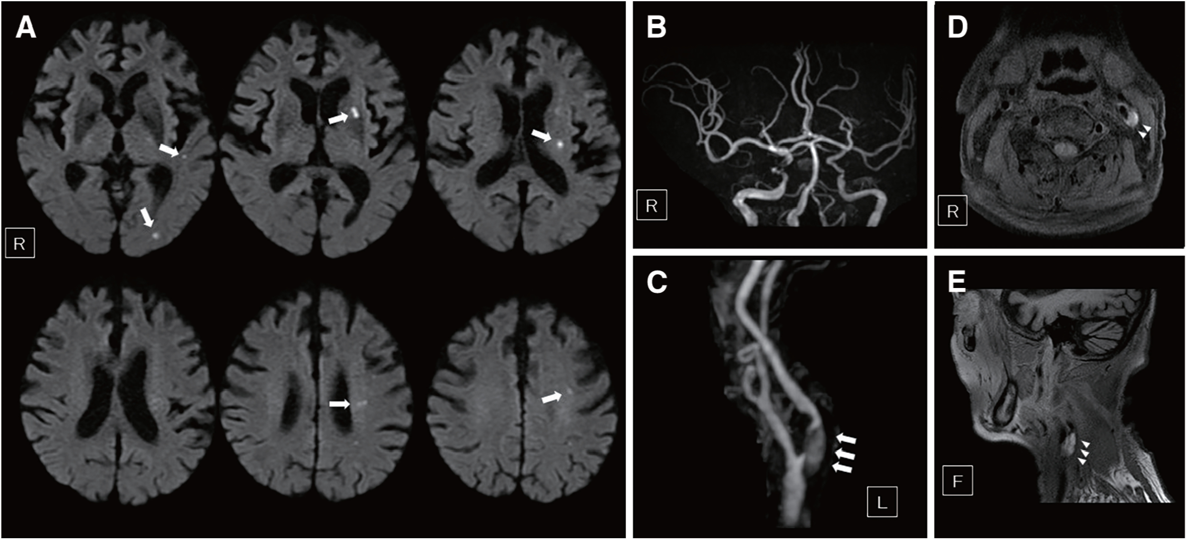
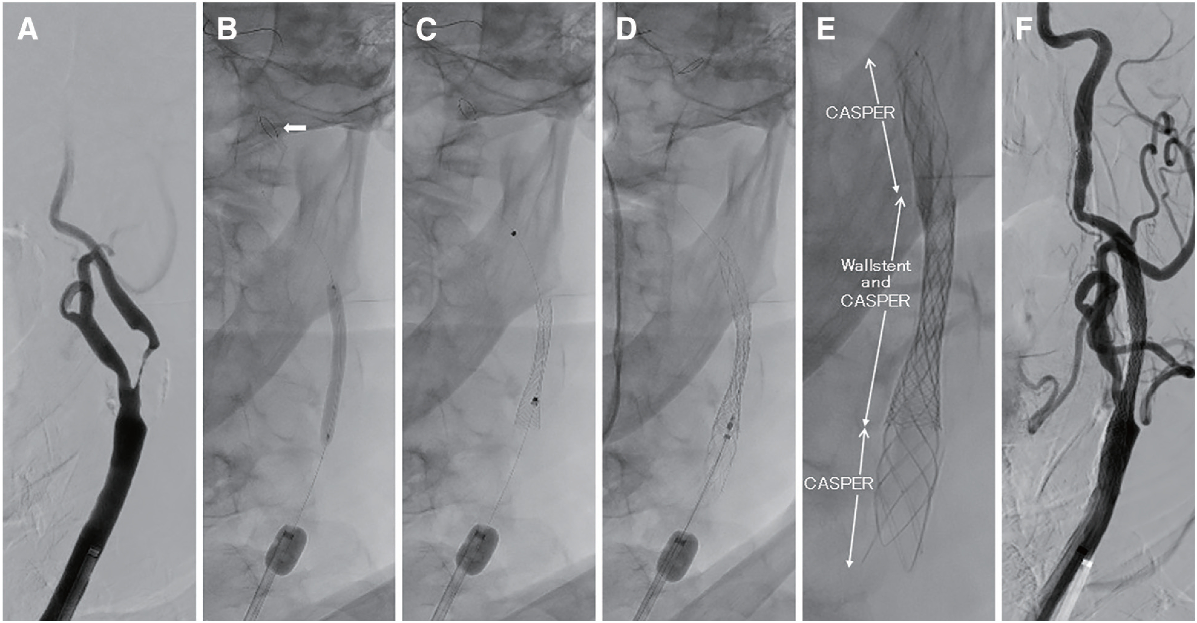
Case presentation: An 87-year-old Japanese man with dysarthria and right hemiparesis was diagnosed with atheromatous cerebral embolism caused by severe left ICA stenosis on MRI and DSA. MRI with T1-weighted black blood methods showed high intensities in the plaques of the left ICA, suggesting unstable plaque characteristics with intraplaque hemorrhage components. On day 20, CAS was performed. After the pre-stent dilation under proximal and distal protection, a Carotid WALLSTENT was placed to cover the stenotic lesion. Then, a CASPER Rx was placed from the proximal left ICA to the common carotid artery to cover the Carotid WALLSTENT. Although visible plaque debris was recognized in the aspirated blood, the debris became invisible after aspiration of 1300 mL. Postoperative angiography showed enough dilation of the left ICA, with no plaque protrusion or acute stent thrombosis. The patient had an uneventful postoperative course and was discharged without any neurological sequelae.
Conclusion: The present case suggests that the combined stent-in-stent technique using a closed-cell stent and a micromesh stent can be considered as one of the treatment strategies for preventing plaque protrusion and procedural ischemic complications in patients with high-risk carotid plaques.


 求助内容:
求助内容: 应助结果提醒方式:


