Martin-Leo Hansmann , Frederick Klauschen , Wojciech Samek , Klaus-Robert Müller , Emmanuel Donnadieu , Sonja Scharf , Sylvia Hartmann , Ina Koch , Jörg Ackermann , Liron Pantanowitz , Hendrik Schäfer , Patrick Wurzel
{"title":"Imaging bridges pathology and radiology","authors":"Martin-Leo Hansmann , Frederick Klauschen , Wojciech Samek , Klaus-Robert Müller , Emmanuel Donnadieu , Sonja Scharf , Sylvia Hartmann , Ina Koch , Jörg Ackermann , Liron Pantanowitz , Hendrik Schäfer , Patrick Wurzel","doi":"10.1016/j.jpi.2023.100298","DOIUrl":null,"url":null,"abstract":"<div><p>In recent years, medical disciplines have moved closer together and rigid borders have been increasingly dissolved. The synergetic advantage of combining multiple disciplines is particularly important for radiology, nuclear medicine, and pathology to perform integrative diagnostics. In this review, we discuss how medical subdisciplines can be reintegrated in the future using state-of-the-art methods of digitization, data science, and machine learning. Integration of methods is made possible by the digitalization of radiological and nuclear medical images, as well as pathological images. 3D histology can become a valuable tool, not only for integration into radiological images but also for the visualization of cellular interactions, the so-called connectomes. In human pathology, it has recently become possible to image and calculate the movements and contacts of immunostained cells in fresh tissue explants. Recording the movement of a living cell is proving to be informative and makes it possible to study dynamic connectomes in the diagnosis of lymphoid tissue. By applying computational methods including data science and machine learning, new perspectives for analyzing and understanding diseases become possible.</p></div>","PeriodicalId":37769,"journal":{"name":"Journal of Pathology Informatics","volume":"14 ","pages":"Article 100298"},"PeriodicalIF":0.0000,"publicationDate":"2023-01-01","publicationTypes":"Journal Article","fieldsOfStudy":null,"isOpenAccess":false,"openAccessPdf":"https://ftp.ncbi.nlm.nih.gov/pub/pmc/oa_pdf/b9/5d/main.PMC9958472.pdf","citationCount":"0","resultStr":null,"platform":"Semanticscholar","paperid":null,"PeriodicalName":"Journal of Pathology Informatics","FirstCategoryId":"1085","ListUrlMain":"https://www.sciencedirect.com/science/article/pii/S2153353923001128","RegionNum":0,"RegionCategory":null,"ArticlePicture":[],"TitleCN":null,"AbstractTextCN":null,"PMCID":null,"EPubDate":"","PubModel":"","JCR":"Q2","JCRName":"Medicine","Score":null,"Total":0}
引用次数: 0
Abstract
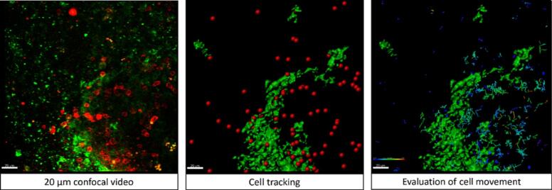
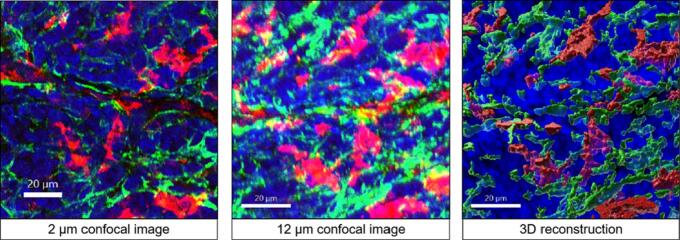
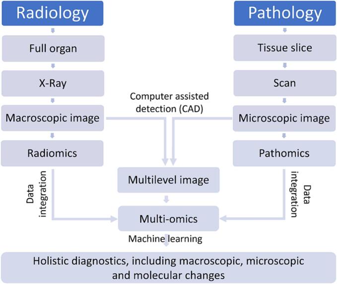
In recent years, medical disciplines have moved closer together and rigid borders have been increasingly dissolved. The synergetic advantage of combining multiple disciplines is particularly important for radiology, nuclear medicine, and pathology to perform integrative diagnostics. In this review, we discuss how medical subdisciplines can be reintegrated in the future using state-of-the-art methods of digitization, data science, and machine learning. Integration of methods is made possible by the digitalization of radiological and nuclear medical images, as well as pathological images. 3D histology can become a valuable tool, not only for integration into radiological images but also for the visualization of cellular interactions, the so-called connectomes. In human pathology, it has recently become possible to image and calculate the movements and contacts of immunostained cells in fresh tissue explants. Recording the movement of a living cell is proving to be informative and makes it possible to study dynamic connectomes in the diagnosis of lymphoid tissue. By applying computational methods including data science and machine learning, new perspectives for analyzing and understanding diseases become possible.
期刊介绍:
The Journal of Pathology Informatics (JPI) is an open access peer-reviewed journal dedicated to the advancement of pathology informatics. This is the official journal of the Association for Pathology Informatics (API). The journal aims to publish broadly about pathology informatics and freely disseminate all articles worldwide. This journal is of interest to pathologists, informaticians, academics, researchers, health IT specialists, information officers, IT staff, vendors, and anyone with an interest in informatics. We encourage submissions from anyone with an interest in the field of pathology informatics. We publish all types of papers related to pathology informatics including original research articles, technical notes, reviews, viewpoints, commentaries, editorials, symposia, meeting abstracts, book reviews, and correspondence to the editors. All submissions are subject to rigorous peer review by the well-regarded editorial board and by expert referees in appropriate specialties.

 求助内容:
求助内容: 应助结果提醒方式:


