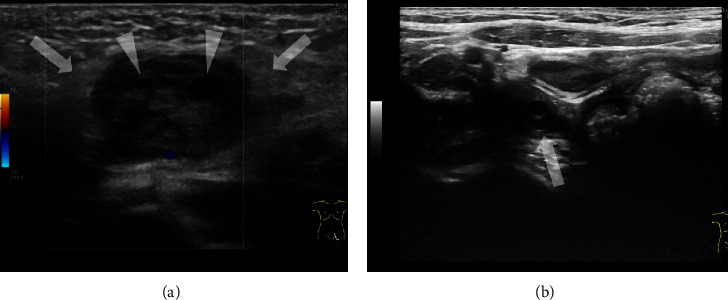
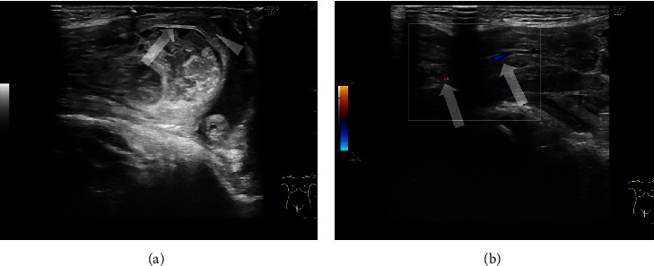
{"title":"Point-of-Care Ultrasound for Mimicker Lesions of Incarcerated Inguinal Hernia.","authors":"Takahiro Hosokawa, Shinsuke Yoshizawa, Kyoichi Deie, Kensuke Ohashi, Hiroshi Kawashima","doi":"10.1155/2023/5593369","DOIUrl":null,"url":null,"abstract":"<p><p>Inguinal hernia is the most common surgical disease in pediatric patients, and urgent intervention such as manual reduction is needed for incarcerated inguinal hernia. Torsion of undescended testes, inguinal herniated ovarian torsion, and purulent lymphadenitis are mimickers of this condition. If these mimicker lesions are misdiagnosed as incarcerated inguinal hernia, manual reduction usually fails, and edematous and erythematous changes may occur in these mimicker lesions due to manual reduction. For physicians in the emergency department, prompt decisions and familiarity with the sonographic appearance of different contents within an inguinal hernia are important to accurately diagnose these mimickers. In this case series, we present sonographic images of a typical case of incarcerated inguinal hernia (an 11-month-old male with right incarcerated inguinal hernia) and three cases of mimicker lesions (a 7-month-old female with herniated ovarian torsion, a 7-year-old boy with undescended testicular torsion, and a 2-month-old male with purulent lymphadenitis). The incidence of incarcerated inguinal hernia is reported to be higher in males (80%), on the right side (60%), and in infants and toddlers. This information is important for diagnosing mimicker lesions. In addition, to prevent manual reduction in mimicker diseases, point-of-care ultrasound before manual reduction in suspected cases of incarcerated inguinal hernia is important.</p>","PeriodicalId":9623,"journal":{"name":"Case Reports in Pediatrics","volume":"2023 ","pages":"5593369"},"PeriodicalIF":0.5000,"publicationDate":"2023-01-01","publicationTypes":"Journal Article","fieldsOfStudy":null,"isOpenAccess":false,"openAccessPdf":"https://www.ncbi.nlm.nih.gov/pmc/articles/PMC10499529/pdf/","citationCount":"0","resultStr":null,"platform":"Semanticscholar","paperid":null,"PeriodicalName":"Case Reports in Pediatrics","FirstCategoryId":"1085","ListUrlMain":"https://doi.org/10.1155/2023/5593369","RegionNum":0,"RegionCategory":null,"ArticlePicture":[],"TitleCN":null,"AbstractTextCN":null,"PMCID":null,"EPubDate":"","PubModel":"","JCR":"Q4","JCRName":"PEDIATRICS","Score":null,"Total":0}
引用次数: 0
Abstract
Inguinal hernia is the most common surgical disease in pediatric patients, and urgent intervention such as manual reduction is needed for incarcerated inguinal hernia. Torsion of undescended testes, inguinal herniated ovarian torsion, and purulent lymphadenitis are mimickers of this condition. If these mimicker lesions are misdiagnosed as incarcerated inguinal hernia, manual reduction usually fails, and edematous and erythematous changes may occur in these mimicker lesions due to manual reduction. For physicians in the emergency department, prompt decisions and familiarity with the sonographic appearance of different contents within an inguinal hernia are important to accurately diagnose these mimickers. In this case series, we present sonographic images of a typical case of incarcerated inguinal hernia (an 11-month-old male with right incarcerated inguinal hernia) and three cases of mimicker lesions (a 7-month-old female with herniated ovarian torsion, a 7-year-old boy with undescended testicular torsion, and a 2-month-old male with purulent lymphadenitis). The incidence of incarcerated inguinal hernia is reported to be higher in males (80%), on the right side (60%), and in infants and toddlers. This information is important for diagnosing mimicker lesions. In addition, to prevent manual reduction in mimicker diseases, point-of-care ultrasound before manual reduction in suspected cases of incarcerated inguinal hernia is important.


 求助内容:
求助内容: 应助结果提醒方式:


