Ceren Aktuna-Belgin, Gozde Serindere, Huseyin Berkay Belgin, Mehmet Serindere, Kaan Orhan
{"title":"Efficacy of low dose and ultra-low dose on the visibility of peri-implant fenestration and dehiscences: a computed tomography study.","authors":"Ceren Aktuna-Belgin, Gozde Serindere, Huseyin Berkay Belgin, Mehmet Serindere, Kaan Orhan","doi":"10.5114/pjr.2022.112466","DOIUrl":null,"url":null,"abstract":"<p><strong>Purpose: </strong>This study aimed to evaluate the visibility of peri-implant fenestration and dehiscences on computed tomography (CT) images taken with 2 different doses.</p><p><strong>Material and methods: </strong>The defects were created on the apical of 6 implants randomly selected from 20 titanium implants placed in the ribs, and dehiscences were created on the cervical of 8 implants. No defects were created around 6 implants. Macroscopic analysis of the implanted ribs was accepted as the gold standard. From the samples, images were taken by using both ultra-low dose (80 kVp, 50 mA, 1.25 mm slice thickness) and low dose (100 kVp, 50 mA, 1.25 mm slice thickness) protocols in CT. The images obtained were evaluated using a 5-point scale.</p><p><strong>Results: </strong>No significant difference was found between the area under the receiver operating characteristic of ultra-low dose protocol and low dose protocol in both defects based on the Wilcoxon test (<i>p</i> > 0.05).</p><p><strong>Conclusions: </strong>The ultra-low dose protocol could be applied by adhering to the \"as low as reasonably achievable\" principle in the diagnosis of peri-implant defects.</p>","PeriodicalId":47128,"journal":{"name":"Polish Journal of Radiology","volume":"87 ","pages":"e24-e29"},"PeriodicalIF":0.9000,"publicationDate":"2022-01-01","publicationTypes":"Journal Article","fieldsOfStudy":null,"isOpenAccess":false,"openAccessPdf":"https://ftp.ncbi.nlm.nih.gov/pub/pmc/oa_pdf/1e/2b/PJR-87-46142.PMC8814895.pdf","citationCount":"1","resultStr":null,"platform":"Semanticscholar","paperid":null,"PeriodicalName":"Polish Journal of Radiology","FirstCategoryId":"1085","ListUrlMain":"https://doi.org/10.5114/pjr.2022.112466","RegionNum":0,"RegionCategory":null,"ArticlePicture":[],"TitleCN":null,"AbstractTextCN":null,"PMCID":null,"EPubDate":"","PubModel":"","JCR":"Q4","JCRName":"RADIOLOGY, NUCLEAR MEDICINE & MEDICAL IMAGING","Score":null,"Total":0}
引用次数: 1
Abstract
Purpose: This study aimed to evaluate the visibility of peri-implant fenestration and dehiscences on computed tomography (CT) images taken with 2 different doses.
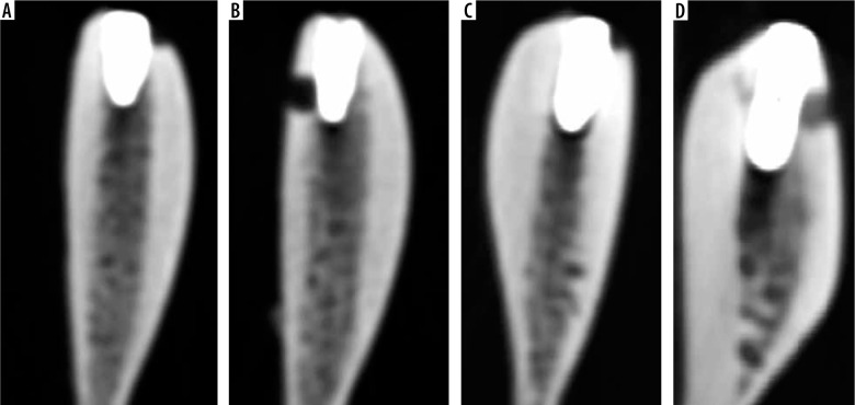
Material and methods: The defects were created on the apical of 6 implants randomly selected from 20 titanium implants placed in the ribs, and dehiscences were created on the cervical of 8 implants. No defects were created around 6 implants. Macroscopic analysis of the implanted ribs was accepted as the gold standard. From the samples, images were taken by using both ultra-low dose (80 kVp, 50 mA, 1.25 mm slice thickness) and low dose (100 kVp, 50 mA, 1.25 mm slice thickness) protocols in CT. The images obtained were evaluated using a 5-point scale.
Results: No significant difference was found between the area under the receiver operating characteristic of ultra-low dose protocol and low dose protocol in both defects based on the Wilcoxon test (p > 0.05).
Conclusions: The ultra-low dose protocol could be applied by adhering to the "as low as reasonably achievable" principle in the diagnosis of peri-implant defects.

 求助内容:
求助内容: 应助结果提醒方式:


