Multi-view carotid ultrasound is stronger associated with cardiovascular risk factors than presence of plaque or single carotid intima media thickness measurements in subclinical atherosclerosis.
Anna Bengtsson, Emma Nyman, Christer Grönlund, Per Wester, Ulf Näslund, Eva Fhärm, Margareta Norberg
{"title":"Multi-view carotid ultrasound is stronger associated with cardiovascular risk factors than presence of plaque or single carotid intima media thickness measurements in subclinical atherosclerosis.","authors":"Anna Bengtsson, Emma Nyman, Christer Grönlund, Per Wester, Ulf Näslund, Eva Fhärm, Margareta Norberg","doi":"10.1007/s10554-023-02868-0","DOIUrl":null,"url":null,"abstract":"<p><p>We aimed to explore the prevalence of atherosclerosis by using multi-view ultrasound examination of the carotid arteries and its association with clinical risk factors in a middle-aged population at low to intermediate risk of cardiovascular disease (CVD). Carotid vascular ultrasound was performed in 3532 participants in the VIPVIZA trial. Mean and maximal carotid intima media thickness (cIMT) at prespecified angles and plaque presence were examined on the left and right side. Associations between CVD risk factors and ultrasound variables were quantified by partial least squares (PLS) regression. A combined ultrasound variable was computed using weights of the first PLS component. Associations between CVD risk factors and the combined multi-view ultrasound variable, single cIMT and plaque measurements, respectively, were determined using linear regression modelling. The participants' mean age was 55.7 years and 52.9% were women. Plaque prevalence was 51.1% in men and 39.0% in women. cIMT was higher in men than in women and in the left compared with the right carotid artery. The strongest association of CVD risk factors was observed with the combined multi-view ultrasound variable (R2 = 24%), compared with single cIMT variables (R2 = 14-18%) and plaque presence (R2 = 15%). The pattern was similar in both sexes. The association with CVD risk factors and the combined ultrasound variable was stronger in 40-year olds (R2 = 22%) compared with 50- or 60-year olds (R = 12%). CVD risk factors are stronger associated with a combined ultrasound variable than plaque presence or single cIMT measures suggesting that carotid multi-view ultrasonography better captures the focality of early atherosclerosis.Clinical Trial Registration: ClinicalTrials.gov, number NCT01849575. May 8, 2013.</p>","PeriodicalId":50332,"journal":{"name":"International Journal of Cardiovascular Imaging","volume":"39 8","pages":"1461-1471"},"PeriodicalIF":1.5000,"publicationDate":"2023-08-01","publicationTypes":"Journal Article","fieldsOfStudy":null,"isOpenAccess":false,"openAccessPdf":"https://www.ncbi.nlm.nih.gov/pmc/articles/PMC10427531/pdf/","citationCount":"1","resultStr":null,"platform":"Semanticscholar","paperid":null,"PeriodicalName":"International Journal of Cardiovascular Imaging","FirstCategoryId":"3","ListUrlMain":"https://doi.org/10.1007/s10554-023-02868-0","RegionNum":4,"RegionCategory":"医学","ArticlePicture":[],"TitleCN":null,"AbstractTextCN":null,"PMCID":null,"EPubDate":"","PubModel":"","JCR":"Q3","JCRName":"CARDIAC & CARDIOVASCULAR SYSTEMS","Score":null,"Total":0}
引用次数: 1
Abstract
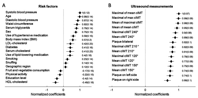
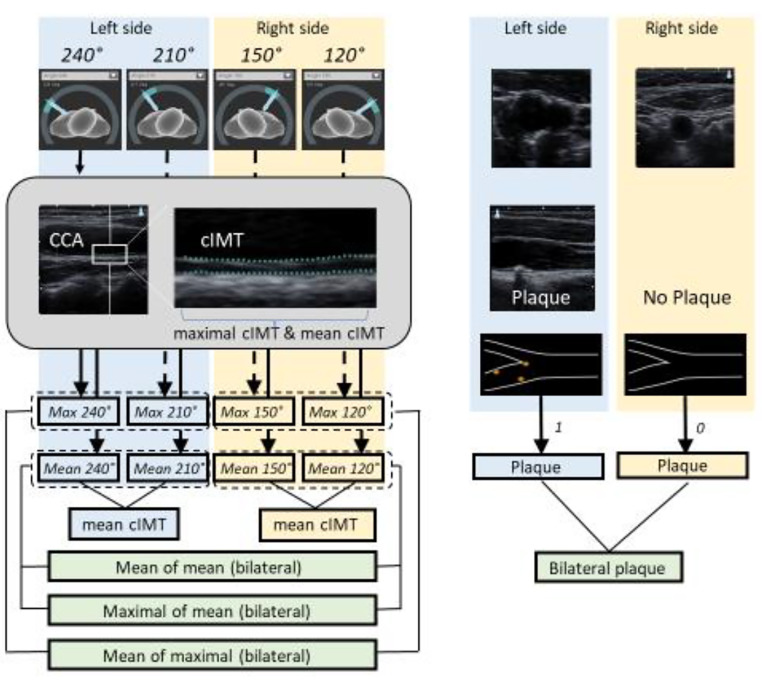
We aimed to explore the prevalence of atherosclerosis by using multi-view ultrasound examination of the carotid arteries and its association with clinical risk factors in a middle-aged population at low to intermediate risk of cardiovascular disease (CVD). Carotid vascular ultrasound was performed in 3532 participants in the VIPVIZA trial. Mean and maximal carotid intima media thickness (cIMT) at prespecified angles and plaque presence were examined on the left and right side. Associations between CVD risk factors and ultrasound variables were quantified by partial least squares (PLS) regression. A combined ultrasound variable was computed using weights of the first PLS component. Associations between CVD risk factors and the combined multi-view ultrasound variable, single cIMT and plaque measurements, respectively, were determined using linear regression modelling. The participants' mean age was 55.7 years and 52.9% were women. Plaque prevalence was 51.1% in men and 39.0% in women. cIMT was higher in men than in women and in the left compared with the right carotid artery. The strongest association of CVD risk factors was observed with the combined multi-view ultrasound variable (R2 = 24%), compared with single cIMT variables (R2 = 14-18%) and plaque presence (R2 = 15%). The pattern was similar in both sexes. The association with CVD risk factors and the combined ultrasound variable was stronger in 40-year olds (R2 = 22%) compared with 50- or 60-year olds (R = 12%). CVD risk factors are stronger associated with a combined ultrasound variable than plaque presence or single cIMT measures suggesting that carotid multi-view ultrasonography better captures the focality of early atherosclerosis.Clinical Trial Registration: ClinicalTrials.gov, number NCT01849575. May 8, 2013.
期刊介绍:
The International Journal of Cardiovascular Imaging publishes technical and clinical communications (original articles, review articles and editorial comments) associated with cardiovascular diseases. The technical communications include the research, development and evaluation of novel imaging methods in the various imaging domains. These domains include magnetic resonance imaging, computed tomography, X-ray imaging, intravascular imaging, and applications in nuclear cardiology and echocardiography, and any combination of these techniques. Of particular interest are topics in medical image processing and image-guided interventions. Clinical applications of such imaging techniques include improved diagnostic approaches, treatment , prognosis and follow-up of cardiovascular patients. Topics include: multi-center or larger individual studies dealing with risk stratification and imaging utilization, applications for better characterization of cardiovascular diseases, and assessment of the efficacy of new drugs and interventional devices.
 求助内容:
求助内容: 应助结果提醒方式:


