Brandon H Schwartz, Balaji K Tamarappoo, Hezzy Shmueli, Robert J Siegel
{"title":"Soft tissue displacement for detection of left ventricle apical dyskinesis with transthoracic echocardiography.","authors":"Brandon H Schwartz, Balaji K Tamarappoo, Hezzy Shmueli, Robert J Siegel","doi":"10.1007/s10554-023-02856-4","DOIUrl":null,"url":null,"abstract":"<p><p>We tested the hypothesis that the use of outward displacement of the soft tissue between the apex and the chest wall as seen in TTE, is a sign of apical displacement and would allow for more accurate diagnosis of apical dyskinesis. This is a retrospective study of 123 patients who underwent TTE and cardiac magnetic resonance imaging (MRI) within a time frame of 6 months between 2008 and 2019. 110 subjects were deemed to have good quality studies and included in the final analysis. An observer blinded to the study objectives evaluated the echocardiograms and recorded the presence or absence of apical dyskinesis. Two independent observers evaluated the echocardiograms based on the presence or absence of outward displacement of the overlying tissue at the LV apex. Cardiac MRI was used to validate the presence of apical dyskinesis. The proportion of studies which were identified as having apical dyskinesis with conventional criteria defined as outward movement of the left ventricular apex during systole were compared to those deemed to have dyskinesis based on tissue displacement. By cardiac MRI, 90 patients had apical dyskinesis. Using conventional criteria on TTE interpretation, 21 were diagnosed with apical dyskinesis (23.3%). However, when soft tissue displacement was used as the diagnostic marker of dyskinesis, 78 patients (86.7%) were diagnosed with dyskinesis, p < 0.01. Detection of displacement of soft tissue overlying the LV apex facilitates better recognition of LV apical dyskinesis.</p>","PeriodicalId":50332,"journal":{"name":"International Journal of Cardiovascular Imaging","volume":"39 8","pages":"1425-1430"},"PeriodicalIF":1.5000,"publicationDate":"2023-08-01","publicationTypes":"Journal Article","fieldsOfStudy":null,"isOpenAccess":false,"openAccessPdf":"https://www.ncbi.nlm.nih.gov/pmc/articles/PMC10427534/pdf/","citationCount":"0","resultStr":null,"platform":"Semanticscholar","paperid":null,"PeriodicalName":"International Journal of Cardiovascular Imaging","FirstCategoryId":"3","ListUrlMain":"https://doi.org/10.1007/s10554-023-02856-4","RegionNum":4,"RegionCategory":"医学","ArticlePicture":[],"TitleCN":null,"AbstractTextCN":null,"PMCID":null,"EPubDate":"","PubModel":"","JCR":"Q3","JCRName":"CARDIAC & CARDIOVASCULAR SYSTEMS","Score":null,"Total":0}
引用次数: 0
Abstract
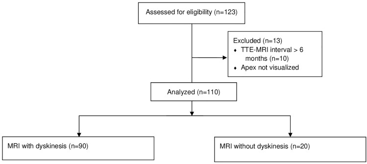
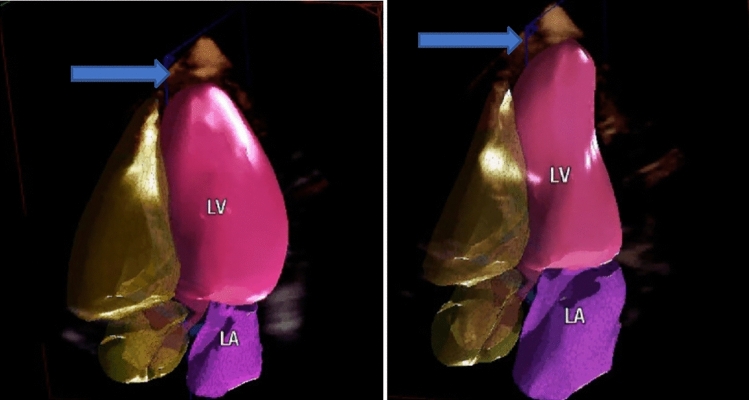
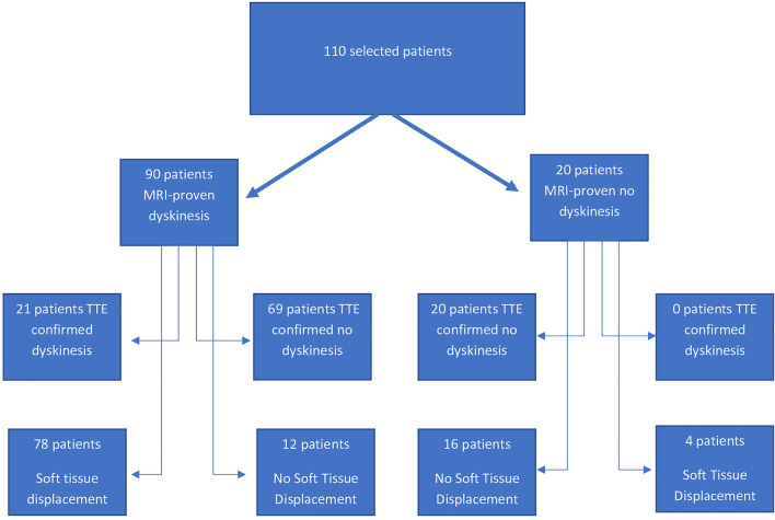
We tested the hypothesis that the use of outward displacement of the soft tissue between the apex and the chest wall as seen in TTE, is a sign of apical displacement and would allow for more accurate diagnosis of apical dyskinesis. This is a retrospective study of 123 patients who underwent TTE and cardiac magnetic resonance imaging (MRI) within a time frame of 6 months between 2008 and 2019. 110 subjects were deemed to have good quality studies and included in the final analysis. An observer blinded to the study objectives evaluated the echocardiograms and recorded the presence or absence of apical dyskinesis. Two independent observers evaluated the echocardiograms based on the presence or absence of outward displacement of the overlying tissue at the LV apex. Cardiac MRI was used to validate the presence of apical dyskinesis. The proportion of studies which were identified as having apical dyskinesis with conventional criteria defined as outward movement of the left ventricular apex during systole were compared to those deemed to have dyskinesis based on tissue displacement. By cardiac MRI, 90 patients had apical dyskinesis. Using conventional criteria on TTE interpretation, 21 were diagnosed with apical dyskinesis (23.3%). However, when soft tissue displacement was used as the diagnostic marker of dyskinesis, 78 patients (86.7%) were diagnosed with dyskinesis, p < 0.01. Detection of displacement of soft tissue overlying the LV apex facilitates better recognition of LV apical dyskinesis.
期刊介绍:
The International Journal of Cardiovascular Imaging publishes technical and clinical communications (original articles, review articles and editorial comments) associated with cardiovascular diseases. The technical communications include the research, development and evaluation of novel imaging methods in the various imaging domains. These domains include magnetic resonance imaging, computed tomography, X-ray imaging, intravascular imaging, and applications in nuclear cardiology and echocardiography, and any combination of these techniques. Of particular interest are topics in medical image processing and image-guided interventions. Clinical applications of such imaging techniques include improved diagnostic approaches, treatment , prognosis and follow-up of cardiovascular patients. Topics include: multi-center or larger individual studies dealing with risk stratification and imaging utilization, applications for better characterization of cardiovascular diseases, and assessment of the efficacy of new drugs and interventional devices.
 求助内容:
求助内容: 应助结果提醒方式:


