Stephen Sai Folmsbee, Douglas R Wilcox, Koen Tyberghein, Pieter De Bleser, Warren G Tourtellotte, Jolanda van Hengel, Frans van Roy, Cara J Gottardi
{"title":"αT-catenin in restricted brain cell types and its potential connection to autism.","authors":"Stephen Sai Folmsbee, Douglas R Wilcox, Koen Tyberghein, Pieter De Bleser, Warren G Tourtellotte, Jolanda van Hengel, Frans van Roy, Cara J Gottardi","doi":"10.1186/s40303-016-0017-9","DOIUrl":null,"url":null,"abstract":"<p><strong>Background: </strong>Recent genetic association studies have linked the cadherin-based adherens junction protein alpha-T-catenin (αT-cat, CTNNA3) with the development of autism. Where αT-cat is expressed in the brain, and how its loss could contribute to this disorder, are entirely unknown.</p><p><strong>Methods: </strong>We used the αT-cat knockout mouse to examine the localization of αT-cat in the brain, and we used histology and immunofluorescence analysis to examine the neurobiological consequences of its loss.</p><p><strong>Results: </strong>We found that αT-cat comprises the ependymal cell junctions of the ventricles of the brain, and its loss led to compensatory upregulation of αE-cat expression. Notably, αT-cat was not detected within the choroid plexus, which relies on cell junction components common to typical epithelial cells. While αT-cat was not detected in neurons of the cerebral cortex, it was abundantly detected within neuronal structures of the molecular layer of the cerebellum. Although αT-cat loss led to no overt differences in cerebral or cerebellar structure, RNA-sequencing analysis from wild type versus knockout cerebella identified a number of disease-relevant signaling pathways associated with αT-cat loss, such as GABA-A receptor activation.</p><p><strong>Conclusions: </strong>These findings raise the possibility that the genetic associations between αT-cat and autism may be due to ependymal and cerebellar defects, and highlight the potential importance of a seemingly redundant adherens junction component to a neurological disorder.</p>","PeriodicalId":73838,"journal":{"name":"Journal of molecular psychiatry","volume":"4 ","pages":"2"},"PeriodicalIF":0.0000,"publicationDate":"2016-06-21","publicationTypes":"Journal Article","fieldsOfStudy":null,"isOpenAccess":false,"openAccessPdf":"https://www.ncbi.nlm.nih.gov/pmc/articles/PMC4915096/pdf/","citationCount":"0","resultStr":null,"platform":"Semanticscholar","paperid":null,"PeriodicalName":"Journal of molecular psychiatry","FirstCategoryId":"1085","ListUrlMain":"https://doi.org/10.1186/s40303-016-0017-9","RegionNum":0,"RegionCategory":null,"ArticlePicture":[],"TitleCN":null,"AbstractTextCN":null,"PMCID":null,"EPubDate":"2016/1/1 0:00:00","PubModel":"eCollection","JCR":"","JCRName":"","Score":null,"Total":0}
引用次数: 0
Abstract
Background: Recent genetic association studies have linked the cadherin-based adherens junction protein alpha-T-catenin (αT-cat, CTNNA3) with the development of autism. Where αT-cat is expressed in the brain, and how its loss could contribute to this disorder, are entirely unknown.
Methods: We used the αT-cat knockout mouse to examine the localization of αT-cat in the brain, and we used histology and immunofluorescence analysis to examine the neurobiological consequences of its loss.
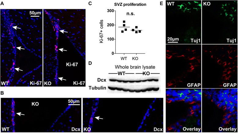
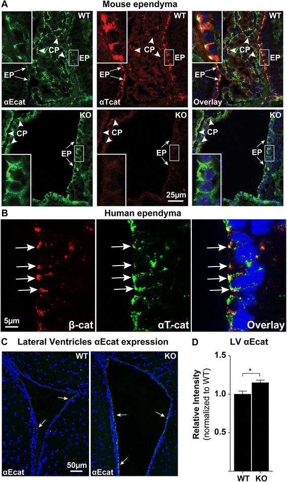
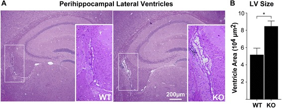
Results: We found that αT-cat comprises the ependymal cell junctions of the ventricles of the brain, and its loss led to compensatory upregulation of αE-cat expression. Notably, αT-cat was not detected within the choroid plexus, which relies on cell junction components common to typical epithelial cells. While αT-cat was not detected in neurons of the cerebral cortex, it was abundantly detected within neuronal structures of the molecular layer of the cerebellum. Although αT-cat loss led to no overt differences in cerebral or cerebellar structure, RNA-sequencing analysis from wild type versus knockout cerebella identified a number of disease-relevant signaling pathways associated with αT-cat loss, such as GABA-A receptor activation.
Conclusions: These findings raise the possibility that the genetic associations between αT-cat and autism may be due to ependymal and cerebellar defects, and highlight the potential importance of a seemingly redundant adherens junction component to a neurological disorder.

 求助内容:
求助内容: 应助结果提醒方式:


