Maria Piagkou, George Tsakotos, George Triantafyllou, Christos Koutserimpas, Dimitrios Chytas, Vasilios Karampelias, Ioannis Pantekidis, Anastasia Triantafyllou, Konstantinos Natsis
{"title":"Coracobrachialis muscle morphology and coexisted neural variants: a cadaveric case series.","authors":"Maria Piagkou, George Tsakotos, George Triantafyllou, Christos Koutserimpas, Dimitrios Chytas, Vasilios Karampelias, Ioannis Pantekidis, Anastasia Triantafyllou, Konstantinos Natsis","doi":"10.1007/s00276-023-03207-7","DOIUrl":null,"url":null,"abstract":"<p><strong>Purpose: </strong>The current cadaveric case series evaluates the coracobrachialis muscle morphology, the related musculocutaneous nerve origin, course, and branching pattern, as well as associated adjacent neuromuscular variants.</p><p><strong>Materials and methods: </strong>Twenty-seven (24 paired and 3 unpaired) cadaveric arms were dissected to identify the coracobrachialis possible variants with emphasis on the musculocutaneous nerve course and coexisted neural variants.</p><p><strong>Results: </strong>Four morphological types of the coracobrachialis were identified: a two-headed muscle in 62.96% (17/27 arms), a three-headed in 22.2% (6/27), a one-headed in 11.1% (3/27), and a four-headed in 3.7% (1 arm). A coracobrachialis variant morphology was identified in 37.04% (10/27). A three-headed biceps brachii muscle coexisted in 23.53% (4/17). Two different courses of the musculocutaneous nerve were recorded: 1. a course between coracobrachialis superficial and deep heads (in cases of two or more heads) (100%, 24/24), and 2. a medial course in case of one-headed coracobrachialis (100%, 3/3). Three neural interconnections were found: 1. the lateral cord of the brachial plexus with the medial root of the median nerve in 18.52%, 2. the musculocutaneous with the median nerve in 7.41% and 3. the radial with the ulnar nerve in 3.71%. Duplication of the lateral root of the median nerve was identified in 11.1%.</p><p><strong>Conclusions: </strong>The knowledge of the morphology of the muscles of the anterior arm compartment, especially the coracobrachialis variant morphology and the related musculocutaneous nerve variable course, is of paramount importance for surgeons. Careful dissection and knowledge of relatively common variants play a significant role in reducing iatrogenic injury.</p>","PeriodicalId":49296,"journal":{"name":"Surgical and Radiologic Anatomy","volume":" ","pages":"1117-1124"},"PeriodicalIF":1.2000,"publicationDate":"2023-09-01","publicationTypes":"Journal Article","fieldsOfStudy":null,"isOpenAccess":false,"openAccessPdf":"https://www.ncbi.nlm.nih.gov/pmc/articles/PMC10514118/pdf/","citationCount":"2","resultStr":null,"platform":"Semanticscholar","paperid":null,"PeriodicalName":"Surgical and Radiologic Anatomy","FirstCategoryId":"3","ListUrlMain":"https://doi.org/10.1007/s00276-023-03207-7","RegionNum":4,"RegionCategory":"医学","ArticlePicture":[],"TitleCN":null,"AbstractTextCN":null,"PMCID":null,"EPubDate":"2023/7/18 0:00:00","PubModel":"Epub","JCR":"Q3","JCRName":"ANATOMY & MORPHOLOGY","Score":null,"Total":0}
引用次数: 2
Abstract
Purpose: The current cadaveric case series evaluates the coracobrachialis muscle morphology, the related musculocutaneous nerve origin, course, and branching pattern, as well as associated adjacent neuromuscular variants.
Materials and methods: Twenty-seven (24 paired and 3 unpaired) cadaveric arms were dissected to identify the coracobrachialis possible variants with emphasis on the musculocutaneous nerve course and coexisted neural variants.
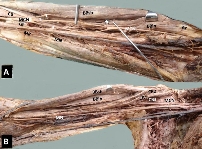
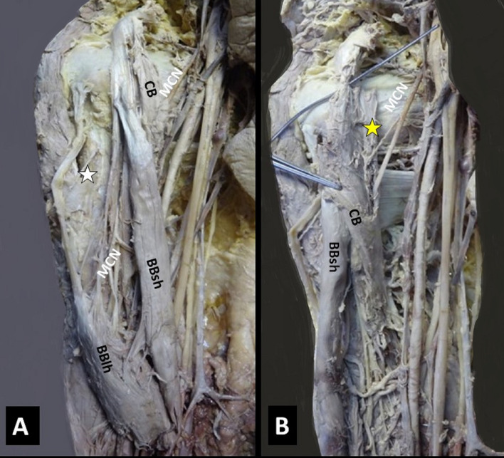
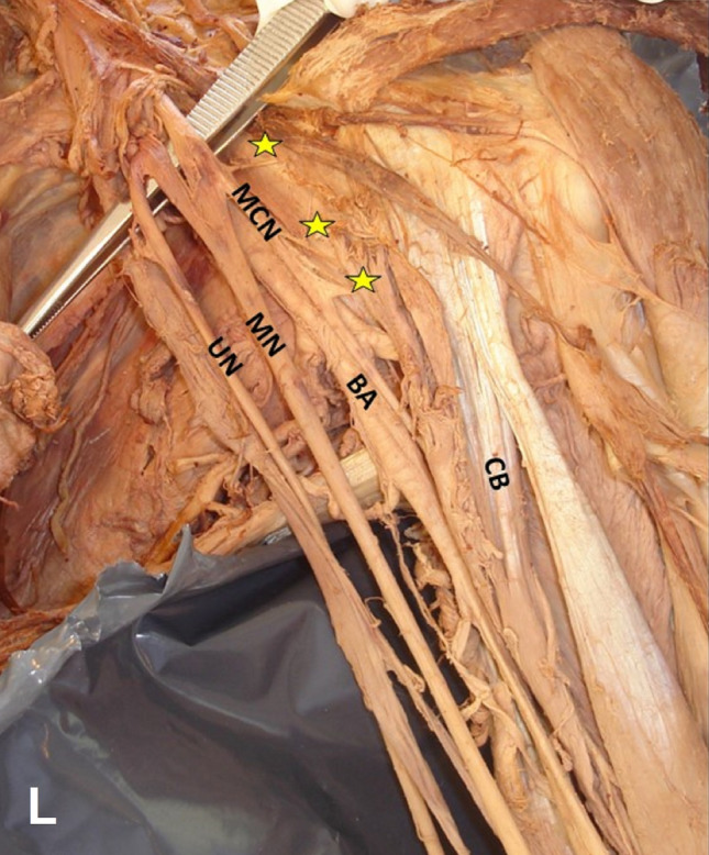
Results: Four morphological types of the coracobrachialis were identified: a two-headed muscle in 62.96% (17/27 arms), a three-headed in 22.2% (6/27), a one-headed in 11.1% (3/27), and a four-headed in 3.7% (1 arm). A coracobrachialis variant morphology was identified in 37.04% (10/27). A three-headed biceps brachii muscle coexisted in 23.53% (4/17). Two different courses of the musculocutaneous nerve were recorded: 1. a course between coracobrachialis superficial and deep heads (in cases of two or more heads) (100%, 24/24), and 2. a medial course in case of one-headed coracobrachialis (100%, 3/3). Three neural interconnections were found: 1. the lateral cord of the brachial plexus with the medial root of the median nerve in 18.52%, 2. the musculocutaneous with the median nerve in 7.41% and 3. the radial with the ulnar nerve in 3.71%. Duplication of the lateral root of the median nerve was identified in 11.1%.
Conclusions: The knowledge of the morphology of the muscles of the anterior arm compartment, especially the coracobrachialis variant morphology and the related musculocutaneous nerve variable course, is of paramount importance for surgeons. Careful dissection and knowledge of relatively common variants play a significant role in reducing iatrogenic injury.
期刊介绍:
Anatomy is a morphological science which cannot fail to interest the clinician. The practical application of anatomical research to clinical problems necessitates special adaptation and selectivity in choosing from numerous international works. Although there is a tendency to believe that meaningful advances in anatomy are unlikely, constant revision is necessary. Surgical and Radiologic Anatomy, the first international journal of Clinical anatomy has been created in this spirit.
Its goal is to serve clinicians, regardless of speciality-physicians, surgeons, radiologists or other specialists-as an indispensable aid with which they can improve their knowledge of anatomy. Each issue includes: Original papers, review articles, articles on the anatomical bases of medical, surgical and radiological techniques, articles of normal radiologic anatomy, brief reviews of anatomical publications of clinical interest.
Particular attention is given to high quality illustrations, which are indispensable for a better understanding of anatomical problems.
Surgical and Radiologic Anatomy is a journal written by anatomists for clinicians with a special interest in anatomy.

 求助内容:
求助内容: 应助结果提醒方式:


