Comparative assessment of surface irregularities of enamel after bonding with different techniques followed by three composite removal methods: An atomic force microscopic study.
Safiya Sana, Mohammed Feroze Hussain, Rony T Kondody, Priyanka Jain
{"title":"Comparative assessment of surface irregularities of enamel after bonding with different techniques followed by three composite removal methods: An atomic force microscopic study.","authors":"Safiya Sana, Mohammed Feroze Hussain, Rony T Kondody, Priyanka Jain","doi":"10.34172/joddd.2023.36867","DOIUrl":null,"url":null,"abstract":"<p><strong>Background: </strong>To compare and assess the enamel surface roughness by Atomic Force Microscopy between ceramic and metal brackets after adhesive removal with 3 different methods.</p><p><strong>Methods: </strong>90 extracted premolars were collected and divided equally into 3 groups G, Y, and R. With group G bonded with metallic brackets (using primer and Transbond XT), group Y with ceramic brackets (primer and Transbond XT), and group R with ceramic brackets (silane and Transbond XT). Each group was subdivided into 3 sub-groups (10 premolars each) based on the resin removal method as A: 12- flute tungsten carbide (TC) bur (high speed), B: 12- flute TC bur (low speed), and C: 30 flute TC bur (low speed). Surface roughness values were calculated and compared before bonding and also after adhesive removal by atomic force microscope (AFM). Measured data were analyzed using paired student t-test, ANOVA, and Tukey's tests.</p><p><strong>Results: </strong>Among the groups, group G showed increased surface roughness after debonding compared to group Y and group R, with Rq value showing a statistically significant difference (<i>P</i><0.047). Whereas, within the subgroups, subgroup A (12-flute TC, high speed) with Rq showed increased surface roughness which was found to be statistically significant (<i>P</i><0.042).</p><p><strong>Conclusion: </strong>None of the adhesive removal methods was capable to restore the enamel to its earlier morphology; a statistically significant increase in surface roughness parameters was reported with a high-speed 12 flute TC bur for Rq and Rt.</p>","PeriodicalId":15599,"journal":{"name":"Journal of Dental Research, Dental Clinics, Dental Prospects","volume":"17 1","pages":"12-17"},"PeriodicalIF":0.0000,"publicationDate":"2023-01-01","publicationTypes":"Journal Article","fieldsOfStudy":null,"isOpenAccess":false,"openAccessPdf":"https://www.ncbi.nlm.nih.gov/pmc/articles/PMC10462917/pdf/","citationCount":"0","resultStr":null,"platform":"Semanticscholar","paperid":null,"PeriodicalName":"Journal of Dental Research, Dental Clinics, Dental Prospects","FirstCategoryId":"1085","ListUrlMain":"https://doi.org/10.34172/joddd.2023.36867","RegionNum":0,"RegionCategory":null,"ArticlePicture":[],"TitleCN":null,"AbstractTextCN":null,"PMCID":null,"EPubDate":"","PubModel":"","JCR":"Q4","JCRName":"Dentistry","Score":null,"Total":0}
引用次数: 0
Abstract
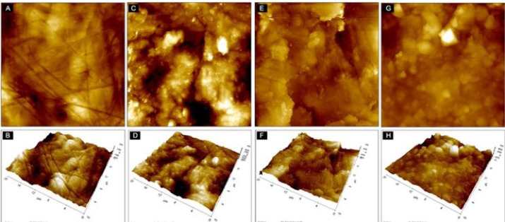
Background: To compare and assess the enamel surface roughness by Atomic Force Microscopy between ceramic and metal brackets after adhesive removal with 3 different methods.
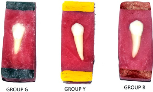
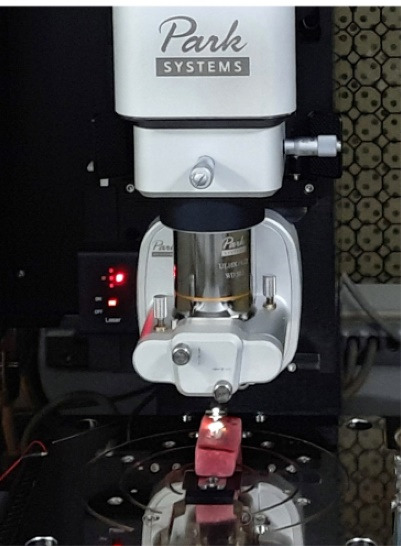
Methods: 90 extracted premolars were collected and divided equally into 3 groups G, Y, and R. With group G bonded with metallic brackets (using primer and Transbond XT), group Y with ceramic brackets (primer and Transbond XT), and group R with ceramic brackets (silane and Transbond XT). Each group was subdivided into 3 sub-groups (10 premolars each) based on the resin removal method as A: 12- flute tungsten carbide (TC) bur (high speed), B: 12- flute TC bur (low speed), and C: 30 flute TC bur (low speed). Surface roughness values were calculated and compared before bonding and also after adhesive removal by atomic force microscope (AFM). Measured data were analyzed using paired student t-test, ANOVA, and Tukey's tests.
Results: Among the groups, group G showed increased surface roughness after debonding compared to group Y and group R, with Rq value showing a statistically significant difference (P<0.047). Whereas, within the subgroups, subgroup A (12-flute TC, high speed) with Rq showed increased surface roughness which was found to be statistically significant (P<0.042).
Conclusion: None of the adhesive removal methods was capable to restore the enamel to its earlier morphology; a statistically significant increase in surface roughness parameters was reported with a high-speed 12 flute TC bur for Rq and Rt.
期刊介绍:
Journal of Dental Research Dental Clinics Dental Prospects (JODDD) is a Platinum* Open Access, peer-reviewed quarterly indexed journal that publishes articles of basic, clinical, and prospective nature in all areas of dentistry and oral health.

 求助内容:
求助内容: 应助结果提醒方式:


