下载PDF
{"title":"Application of CAD/CAM technologies and materials for prosthetic restoration of severely damaged teeth—clinical cases","authors":"Z Tomova, Y Zhekov, G Alexandrov, A Vlahova, E Vasileva","doi":"10.1111/adj.12976","DOIUrl":null,"url":null,"abstract":"<p>In cases of severely damaged teeth with limited coronal tooth structure and remaining hard dental tissues subgingivally, a custom-made post-and-core restoration is required. Teeth with non-circular canal space also require this type of restoration because the build-up with pre-fabricated posts could lead to thick cement layer. The development of CAD/CAM technologies widens the range of the materials that can be used for prosthetic restorations. Along with base dental alloys, newly developed materials may be applied. The aim of the article is to present four clinical cases of severely damaged teeth which utilize different materials and different production techniques for custom post-and-core fabrication. In the first clinical case, a metal post-and-core restoration was fabricated by direct metal laser sintering. In the second clinical case, digital technologies were used to produce a 3D-printed resin prototype for further investing and casting from base metal dental alloy. In the third clinical case, fibre-reinforced composite was used for fabrication of the custom post-and-core by milling. In the fourth clinical case, the restoration is produced by milling of lithium disilicate ceramics IPS emax CAD (Ivoclar Vivadent, Lichtenstein). The bond between the fibre-reinforced composite post-and-core and the hard dental tissues offered possibility to compensate—to some extent—the shape of the preparation which was not optimal. CAD/CAM technologies applied in these clinical cases provided combination of high accuracy of fitting with good stability and individual shape of the restorations. © 2023 Australian Dental Association.</p>","PeriodicalId":8593,"journal":{"name":"Australian dental journal","volume":"68 4","pages":"294-302"},"PeriodicalIF":2.4000,"publicationDate":"2023-09-08","publicationTypes":"Journal Article","fieldsOfStudy":null,"isOpenAccess":false,"openAccessPdf":"https://onlinelibrary.wiley.com/doi/epdf/10.1111/adj.12976","citationCount":"1","resultStr":null,"platform":"Semanticscholar","paperid":null,"PeriodicalName":"Australian dental journal","FirstCategoryId":"3","ListUrlMain":"https://onlinelibrary.wiley.com/doi/10.1111/adj.12976","RegionNum":4,"RegionCategory":"医学","ArticlePicture":[],"TitleCN":null,"AbstractTextCN":null,"PMCID":null,"EPubDate":"","PubModel":"","JCR":"Q2","JCRName":"DENTISTRY, ORAL SURGERY & MEDICINE","Score":null,"Total":0}
引用次数: 1
引用
批量引用
Abstract
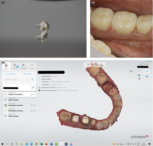
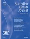
In cases of severely damaged teeth with limited coronal tooth structure and remaining hard dental tissues subgingivally, a custom-made post-and-core restoration is required. Teeth with non-circular canal space also require this type of restoration because the build-up with pre-fabricated posts could lead to thick cement layer. The development of CAD/CAM technologies widens the range of the materials that can be used for prosthetic restorations. Along with base dental alloys, newly developed materials may be applied. The aim of the article is to present four clinical cases of severely damaged teeth which utilize different materials and different production techniques for custom post-and-core fabrication. In the first clinical case, a metal post-and-core restoration was fabricated by direct metal laser sintering. In the second clinical case, digital technologies were used to produce a 3D-printed resin prototype for further investing and casting from base metal dental alloy. In the third clinical case, fibre-reinforced composite was used for fabrication of the custom post-and-core by milling. In the fourth clinical case, the restoration is produced by milling of lithium disilicate ceramics IPS emax CAD (Ivoclar Vivadent, Lichtenstein). The bond between the fibre-reinforced composite post-and-core and the hard dental tissues offered possibility to compensate—to some extent—the shape of the preparation which was not optimal. CAD/CAM technologies applied in these clinical cases provided combination of high accuracy of fitting with good stability and individual shape of the restorations. © 2023 Australian Dental Association.
CAD/CAM技术及材料在重度牙体损伤修复中的应用-临床案例
如果牙齿严重受损,冠状牙结构有限,牙龈下仍有坚硬的牙组织,则需要定制桩核修复。非圆形牙根管空间的牙齿也需要这种类型的修复,因为预制桩的建立可能导致厚厚的水泥层。CAD/CAM技术的发展扩大了可用于假肢修复的材料范围。除了齿科基合金外,还可以应用新开发的材料。本文的目的是介绍四个临床病例严重受损的牙齿,利用不同的材料和不同的生产技术,定制桩核制造。在第一例临床病例中,采用直接金属激光烧结方法制作金属桩核修复体。在第二个临床案例中,数字技术用于生产3d打印树脂原型,用于进一步投资和铸造贱金属牙科合金。在第三个临床病例中,纤维增强复合材料被用于铣削制造定制桩核。在第四个临床病例中,修复是通过研磨二硅酸锂陶瓷IPS emax CAD (Ivoclar Vivadent, Lichtenstein)进行的。纤维增强复合材料桩核与硬牙组织之间的结合在某种程度上提供了补偿制备材料形状不理想的可能性。在这些临床病例中应用的CAD/CAM技术提供了高准确性和良好的稳定性和个性化修复体形状的结合。©2023澳大利亚牙科协会。
本文章由计算机程序翻译,如有差异,请以英文原文为准。
 求助内容:
求助内容: 应助结果提醒方式:


