Mirjam Steiner, Hannah Schwarz, Gregor Kasprian, Judith Rittenschober-Boehm, Victor Schmidbauer, Renate Fuiko, Monika Olischar, Katrin Klebermass-Schrehof, Angelika Berger, Katharina Goeral
{"title":"Brain Biometry Reveals Impaired Brain Growth in Preterm Neonates with Intraventricular Hemorrhage.","authors":"Mirjam Steiner, Hannah Schwarz, Gregor Kasprian, Judith Rittenschober-Boehm, Victor Schmidbauer, Renate Fuiko, Monika Olischar, Katrin Klebermass-Schrehof, Angelika Berger, Katharina Goeral","doi":"10.1159/000528981","DOIUrl":null,"url":null,"abstract":"<p><strong>Introduction: </strong>Preterm birth and cerebral hemorrhage have adverse effects on brain development. Alterations in regional brain size on magnetic resonance imaging (MRI) can be assessed using 2D biometrical analysis, an easily applicable technique showing good correlation with 3D brain volumes.</p><p><strong>Methods: </strong>This retrospective study included 74 preterm neonates with intraventricular hemorrhage (IVH) born <32+0 weeks of gestation between 2011 and 2019. Cerebral MRI was performed at term-equivalent age, and 2D measurement techniques were used for biometrical analysis and compared to normative data of two control groups. Finally, the correlation and association of brain parameters and patterns of impaired brain growth and outcome at 2 and 3 years of age were evaluated.</p><p><strong>Results: </strong>Interhemispheric distance (IHD), the 3rd ventricle, and lateral ventricles presented larger, in contrast, cerebral biparietal width (cBPW), fronto-occipital diameter (FOD), and the length of the corpus callosum were smaller in IVH patients compared to respective controls. The strongest correlations with outcome were observed for the parameters FOD, anteroposterior diameter of the vermis, transverse cerebellar diameter (tCD), corpus callosum, 3rd ventricle, and left ventricular index. Patients with the small FOD, small BPW, and increased IHD pattern reached overall lower outcome scores at follow-up.</p><p><strong>Discussion: </strong>Preterm neonates with IVH showed reduced total brain sizes and enlarged pericerebral spaces compared to neurologically healthy controls. Biometric analysis revealed that several 2D brain parameters as well as different patterns of impaired brain growth were associated with neurodevelopmental impairment in early childhood. These findings may support prediction of long-term outcome and parental counseling in patients with IVH.</p>","PeriodicalId":18924,"journal":{"name":"Neonatology","volume":"120 2","pages":"225-234"},"PeriodicalIF":3.0000,"publicationDate":"2023-01-01","publicationTypes":"Journal Article","fieldsOfStudy":null,"isOpenAccess":false,"openAccessPdf":"https://www.ncbi.nlm.nih.gov/pmc/articles/PMC10906469/pdf/","citationCount":"0","resultStr":null,"platform":"Semanticscholar","paperid":null,"PeriodicalName":"Neonatology","FirstCategoryId":"3","ListUrlMain":"https://doi.org/10.1159/000528981","RegionNum":3,"RegionCategory":"医学","ArticlePicture":[],"TitleCN":null,"AbstractTextCN":null,"PMCID":null,"EPubDate":"2023/2/20 0:00:00","PubModel":"Epub","JCR":"Q1","JCRName":"PEDIATRICS","Score":null,"Total":0}
引用次数: 0
Abstract
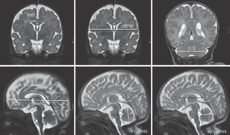
Introduction: Preterm birth and cerebral hemorrhage have adverse effects on brain development. Alterations in regional brain size on magnetic resonance imaging (MRI) can be assessed using 2D biometrical analysis, an easily applicable technique showing good correlation with 3D brain volumes.
Methods: This retrospective study included 74 preterm neonates with intraventricular hemorrhage (IVH) born <32+0 weeks of gestation between 2011 and 2019. Cerebral MRI was performed at term-equivalent age, and 2D measurement techniques were used for biometrical analysis and compared to normative data of two control groups. Finally, the correlation and association of brain parameters and patterns of impaired brain growth and outcome at 2 and 3 years of age were evaluated.
Results: Interhemispheric distance (IHD), the 3rd ventricle, and lateral ventricles presented larger, in contrast, cerebral biparietal width (cBPW), fronto-occipital diameter (FOD), and the length of the corpus callosum were smaller in IVH patients compared to respective controls. The strongest correlations with outcome were observed for the parameters FOD, anteroposterior diameter of the vermis, transverse cerebellar diameter (tCD), corpus callosum, 3rd ventricle, and left ventricular index. Patients with the small FOD, small BPW, and increased IHD pattern reached overall lower outcome scores at follow-up.
Discussion: Preterm neonates with IVH showed reduced total brain sizes and enlarged pericerebral spaces compared to neurologically healthy controls. Biometric analysis revealed that several 2D brain parameters as well as different patterns of impaired brain growth were associated with neurodevelopmental impairment in early childhood. These findings may support prediction of long-term outcome and parental counseling in patients with IVH.
期刊介绍:
This highly respected and frequently cited journal is a prime source of information in the area of fetal and neonatal research. Original papers present research on all aspects of neonatology, fetal medicine and developmental biology. These papers encompass both basic science and clinical research including randomized trials, observational studies and epidemiology. Basic science research covers molecular biology, molecular genetics, physiology, biochemistry and pharmacology in fetal and neonatal life. In addition to the classic features the journal accepts papers for the sections Research Briefings and Sources of Neonatal Medicine (historical pieces). Papers reporting results of animal studies should be based upon hypotheses that relate to developmental processes or disorders in the human fetus or neonate.
 求助内容:
求助内容: 应助结果提醒方式:


