Shalinee Dhayal, Kaiyven Afi Leslie, Mohammad Baity, Pouria Akhbari, Sarah J Richardson, Mark A Russell, Noel G Morgan
{"title":"Temporal regulation of interferon signalling in human EndoC-βH1 cells.","authors":"Shalinee Dhayal, Kaiyven Afi Leslie, Mohammad Baity, Pouria Akhbari, Sarah J Richardson, Mark A Russell, Noel G Morgan","doi":"10.1530/JME-21-0224","DOIUrl":null,"url":null,"abstract":"<p><p>During the development of type 1 diabetes, interferons (IFN) are elaborated from islet-infiltrating immune cells and/or from virally infected β-cells. They act via specific receptors to increase, acutely, the phosphorylation of the transcription factors STAT1 and 2. However, the longer-term impacts of chronic IFN stimulation are poorly understood and were investigated in the current study. Human EndoC-βH1 cells were treated with IFNα, IFNγ or IFNλ either acutely (<2 h) or chronically (≥24 h) and STAT phosphorylation, expression and activity were assessed by Western blotting and transcriptional reporter assays. Exposure of β-cells to IFNα or IFNλ induced a swift increase in the phosphorylation of both STAT1 and STAT2, whereas IFNγ increased only pSTAT1. Over more extended periods (≥24 h), STAT phosphorylation declined but STAT1 and STAT2 expression were enhanced in a sustained manner. All IFNs stimulated ISRE transcriptional activity (but with different time courses), whereas GAS activity was responsive only to IFNγ. The re-addition of a second bolus of IFNα, 24 h after an initial dose, failed to cause renewed STAT1/2 phosphorylation. By contrast, when IFNγ was added 24 h after exposure to IFNα, rapid STAT1 phosphorylation was re-initiated. Exposure of β-cells to IFNs leads to rapid, transient, STAT phosphorylation and to slower and more sustained increases in total STAT1/2 levels. The initial phosphorylation response is accompanied by marked desensitisation to the cognate agonist. Together, the results reveal that the response of β-cells to IFNs is regulated both temporally and quantitatively to achieve effective signal integration.</p>","PeriodicalId":16570,"journal":{"name":"Journal of molecular endocrinology","volume":"69 2","pages":"299-313"},"PeriodicalIF":3.8000,"publicationDate":"2022-05-19","publicationTypes":"Journal Article","fieldsOfStudy":null,"isOpenAccess":false,"openAccessPdf":"https://www.ncbi.nlm.nih.gov/pmc/articles/PMC9175560/pdf/","citationCount":"0","resultStr":null,"platform":"Semanticscholar","paperid":null,"PeriodicalName":"Journal of molecular endocrinology","FirstCategoryId":"3","ListUrlMain":"https://doi.org/10.1530/JME-21-0224","RegionNum":4,"RegionCategory":"医学","ArticlePicture":[],"TitleCN":null,"AbstractTextCN":null,"PMCID":null,"EPubDate":"","PubModel":"","JCR":"Q2","JCRName":"ENDOCRINOLOGY & METABOLISM","Score":null,"Total":0}
引用次数: 0
Abstract
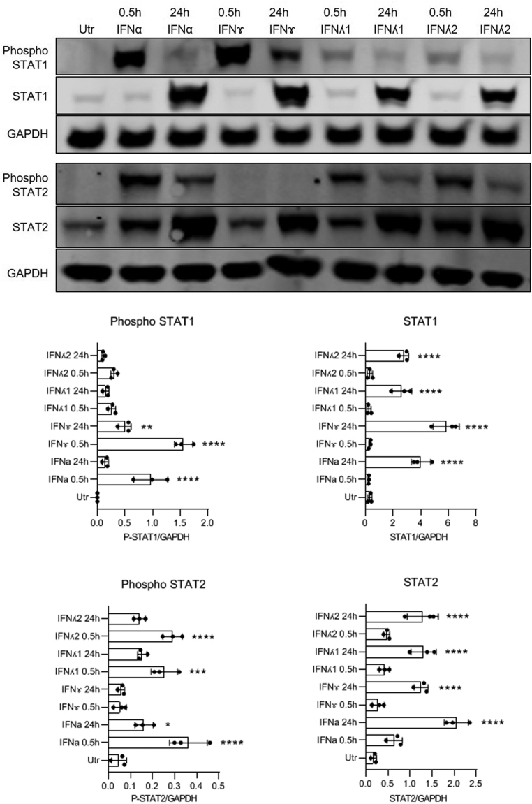
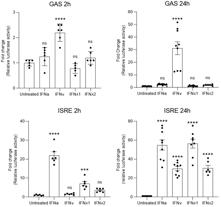
During the development of type 1 diabetes, interferons (IFN) are elaborated from islet-infiltrating immune cells and/or from virally infected β-cells. They act via specific receptors to increase, acutely, the phosphorylation of the transcription factors STAT1 and 2. However, the longer-term impacts of chronic IFN stimulation are poorly understood and were investigated in the current study. Human EndoC-βH1 cells were treated with IFNα, IFNγ or IFNλ either acutely (<2 h) or chronically (≥24 h) and STAT phosphorylation, expression and activity were assessed by Western blotting and transcriptional reporter assays. Exposure of β-cells to IFNα or IFNλ induced a swift increase in the phosphorylation of both STAT1 and STAT2, whereas IFNγ increased only pSTAT1. Over more extended periods (≥24 h), STAT phosphorylation declined but STAT1 and STAT2 expression were enhanced in a sustained manner. All IFNs stimulated ISRE transcriptional activity (but with different time courses), whereas GAS activity was responsive only to IFNγ. The re-addition of a second bolus of IFNα, 24 h after an initial dose, failed to cause renewed STAT1/2 phosphorylation. By contrast, when IFNγ was added 24 h after exposure to IFNα, rapid STAT1 phosphorylation was re-initiated. Exposure of β-cells to IFNs leads to rapid, transient, STAT phosphorylation and to slower and more sustained increases in total STAT1/2 levels. The initial phosphorylation response is accompanied by marked desensitisation to the cognate agonist. Together, the results reveal that the response of β-cells to IFNs is regulated both temporally and quantitatively to achieve effective signal integration.
期刊介绍:
The Journal of Molecular Endocrinology is an official journal of the Society for Endocrinology and is endorsed by the European Society of Endocrinology and the Endocrine Society of Australia.
Journal of Molecular Endocrinology is a leading global journal that publishes original research articles and reviews. The journal focuses on molecular and cellular mechanisms in endocrinology, including: gene regulation, cell biology, signalling, mutations, transgenics, hormone-dependant cancers, nuclear receptors, and omics. Basic and pathophysiological studies at the molecule and cell level are considered, as well as human sample studies where this is the experimental model of choice. Technique studies including CRISPR or gene editing are also encouraged.

 求助内容:
求助内容: 应助结果提醒方式:


