Andrew Tirsi, Paras P Shah, Vasiliki Gliagias, Daniel Barmas-Alamdari, Derek Orshan, Joby Tsai, Celso Tello
{"title":"Posterior Pole Asymmetry Analysis as a Diagnostic Tool in Glaucoma Suspects: An Electrophysiological Approach.","authors":"Andrew Tirsi, Paras P Shah, Vasiliki Gliagias, Daniel Barmas-Alamdari, Derek Orshan, Joby Tsai, Celso Tello","doi":"10.2147/OPTH.S411647","DOIUrl":null,"url":null,"abstract":"<p><strong>Purpose: </strong>Spectral domain optical coherence tomography (SD-OCT) with posterior pole asymmetry analysis (PPAA) provides a mapping of posterior pole retinal thickness with asymmetry analysis between hemispheres of each eye. We investigated whether these structural abnormalities were correlated with functional retinal ganglion cell (RGC) loss, quantified by steady state pattern electroretinogram (ssPERG), in glaucoma suspects (GS).</p><p><strong>Methods: </strong>Twenty GS (34 eyes) were enrolled in a prospective study at the Manhattan Eye, Ear, and Throat Hospital. All subjects underwent ophthalmological examination, including Humphrey visual field, Spectralis Glaucoma Module Premium Edition (GMPE) SD-OCT PPAA, and ssPERG testing. The ability of ssPERG parameters (Magnitude [Mag, µv], MagnitudeD [MagD, µv], and MagD/Mag ratio) to predict PPAA thickness (total, superior, and inferior thickness, [µm]) was tested via adjusted multivariate linear regression analysis.</p><p><strong>Results: </strong>Mag explained 8% of variance in total PPAA change (F(1,29)=6.33, B=6.86, 95% CI: 1.29-12.44, p=0.018), 8% in superior PPAA change (F(1,29)=5.57, B=6.92, 95% CI: 0.92-12.92, p=0.025), and 7.1% in inferior PPAA change (F(1,29)=5.83, B=6.80, 95% CI: 1.04-12.56, p=0.022). Similarly, MagD explained 9.7% of variance in total PPAA change (F(1,29)=8.09, B=6.47, 95% CI: 1.82-11.13, p=0.008), 10% in superior PPAA change (F(1,29)=7.33, B=6.63, 95% CI: 1.62-11.63, p=0.011), and 8.5% in inferior PPAA change (F(1,29)=7.25, B=6.36, 95% CI: 1.53-11.18, p=0.012). MagD/Mag ratio and PPAA were not significantly associated.</p><p><strong>Conclusion: </strong>To the best of our knowledge, this is the first study demonstrating a positive relationship between RGC dysfunction and retinal thickness changes between the superior and inferior hemispheres. The detection of asymmetrical structural loss, combined with functional RGC assessment using ssPERG, may be an informative tool for early glaucoma diagnosis.</p>","PeriodicalId":10442,"journal":{"name":"Clinical ophthalmology","volume":"17 ","pages":"1777-1787"},"PeriodicalIF":1.8000,"publicationDate":"2023-06-21","publicationTypes":"Journal Article","fieldsOfStudy":null,"isOpenAccess":false,"openAccessPdf":"https://ftp.ncbi.nlm.nih.gov/pub/pmc/oa_pdf/82/b4/opth-17-1777.PMC10290849.pdf","citationCount":"0","resultStr":null,"platform":"Semanticscholar","paperid":null,"PeriodicalName":"Clinical ophthalmology","FirstCategoryId":"1085","ListUrlMain":"https://doi.org/10.2147/OPTH.S411647","RegionNum":0,"RegionCategory":null,"ArticlePicture":[],"TitleCN":null,"AbstractTextCN":null,"PMCID":null,"EPubDate":"2023/1/1 0:00:00","PubModel":"eCollection","JCR":"Q3","JCRName":"OPHTHALMOLOGY","Score":null,"Total":0}
引用次数: 0
Abstract
Purpose: Spectral domain optical coherence tomography (SD-OCT) with posterior pole asymmetry analysis (PPAA) provides a mapping of posterior pole retinal thickness with asymmetry analysis between hemispheres of each eye. We investigated whether these structural abnormalities were correlated with functional retinal ganglion cell (RGC) loss, quantified by steady state pattern electroretinogram (ssPERG), in glaucoma suspects (GS).
Methods: Twenty GS (34 eyes) were enrolled in a prospective study at the Manhattan Eye, Ear, and Throat Hospital. All subjects underwent ophthalmological examination, including Humphrey visual field, Spectralis Glaucoma Module Premium Edition (GMPE) SD-OCT PPAA, and ssPERG testing. The ability of ssPERG parameters (Magnitude [Mag, µv], MagnitudeD [MagD, µv], and MagD/Mag ratio) to predict PPAA thickness (total, superior, and inferior thickness, [µm]) was tested via adjusted multivariate linear regression analysis.
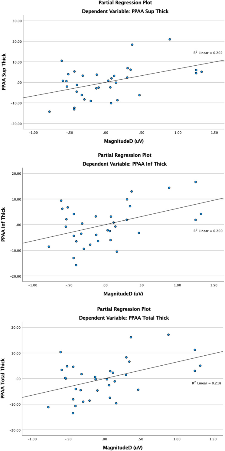
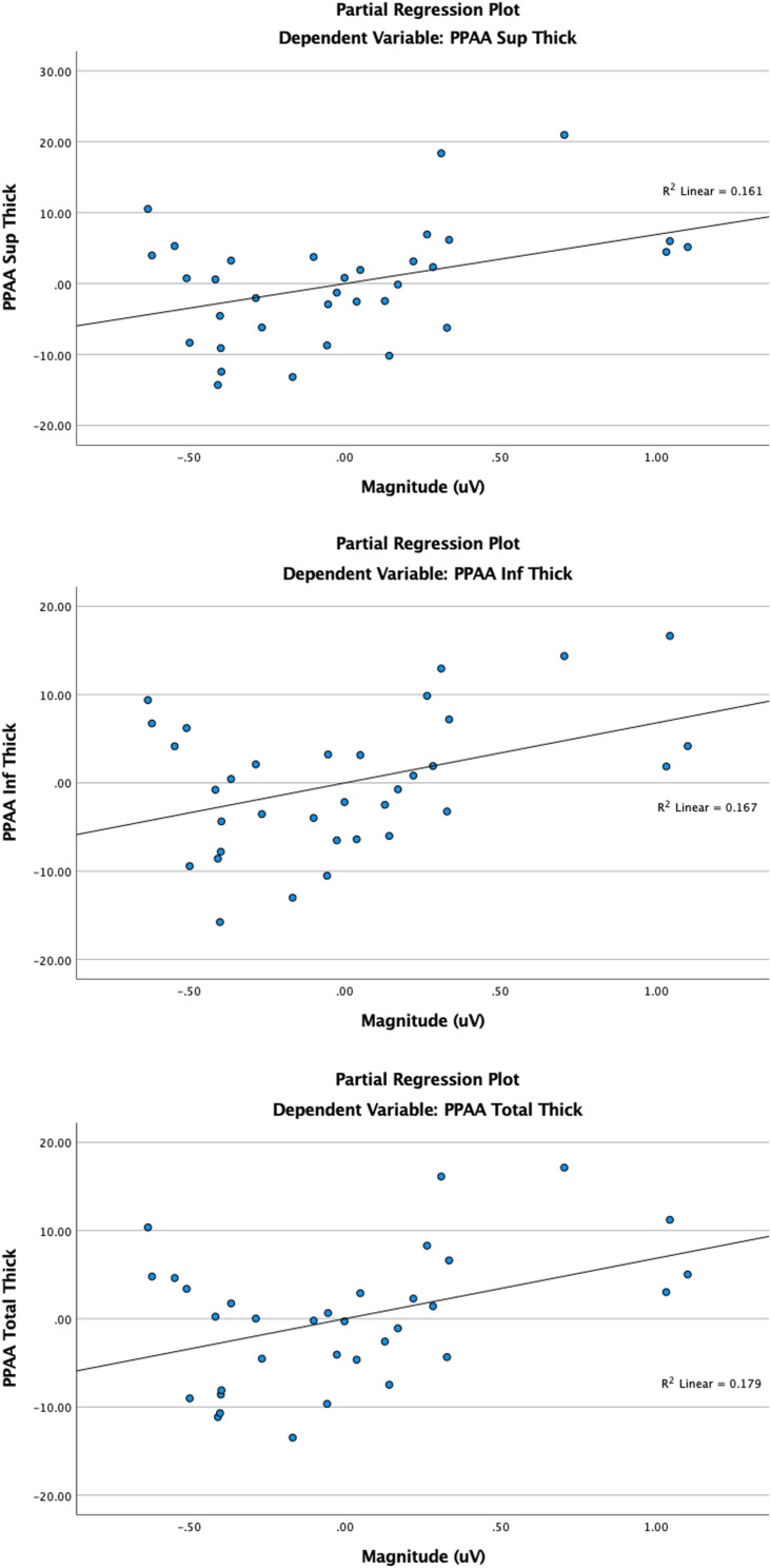
Results: Mag explained 8% of variance in total PPAA change (F(1,29)=6.33, B=6.86, 95% CI: 1.29-12.44, p=0.018), 8% in superior PPAA change (F(1,29)=5.57, B=6.92, 95% CI: 0.92-12.92, p=0.025), and 7.1% in inferior PPAA change (F(1,29)=5.83, B=6.80, 95% CI: 1.04-12.56, p=0.022). Similarly, MagD explained 9.7% of variance in total PPAA change (F(1,29)=8.09, B=6.47, 95% CI: 1.82-11.13, p=0.008), 10% in superior PPAA change (F(1,29)=7.33, B=6.63, 95% CI: 1.62-11.63, p=0.011), and 8.5% in inferior PPAA change (F(1,29)=7.25, B=6.36, 95% CI: 1.53-11.18, p=0.012). MagD/Mag ratio and PPAA were not significantly associated.
Conclusion: To the best of our knowledge, this is the first study demonstrating a positive relationship between RGC dysfunction and retinal thickness changes between the superior and inferior hemispheres. The detection of asymmetrical structural loss, combined with functional RGC assessment using ssPERG, may be an informative tool for early glaucoma diagnosis.

 求助内容:
求助内容: 应助结果提醒方式:


