Zita Reisz, Bence Laszlo Radics, Peter Nemes, Ross Laxton, Laszlo Kaizer, Krisztina Mita Gabor, Timea Novak, Pal Barzo, Safa Al-Sarraj, Istvan Bodi
{"title":"Case Report: Brainstem angiocentric glioma presenting in a toddler child-diagnostic and therapeutic challenges.","authors":"Zita Reisz, Bence Laszlo Radics, Peter Nemes, Ross Laxton, Laszlo Kaizer, Krisztina Mita Gabor, Timea Novak, Pal Barzo, Safa Al-Sarraj, Istvan Bodi","doi":"10.3389/pore.2023.1611231","DOIUrl":null,"url":null,"abstract":"<p><p><b>Introduction:</b> Angiocentric gliomas (AG) in brainstem location are exceedingly rare and might cause differential diagnostic problems and uncertainty regarding the best therapeutic approach. Hereby, we describe the clinicopathological findings in a brainstem AG presenting in a toddler child and review the literature. <b>Case report:</b> A 2-year-old boy presented with 5 weeks history of gait disturbances, frequent falls, left-sided torticollis and swallowing problems. MRI head showed a T2-hyperintense, partly exophytic mass lesion centred in the pontomedullary region, raising the possibility of diffuse midline glioma. The exophytic component was partially resected by suboccipital craniotomy, leaving intact the infiltrative component. Ventriculoperitoneal shunt was implanted due to postoperative hydrocephalus. Histological examination revealed a moderately cellular tumour consisted of bland glial cells infiltrating the brain parenchyma and radially arranged around the blood vessels. By immunohistochemistry, the tumour strongly expressed S100 and GFAP in addition to intense nestin positivity, while OLIG2 was negative in the perivascular tumour cells. DNA methylation array profiled the tumour as \"methylation class diffuse astrocytoma, <i>MYB</i> or <i>MYBL1</i>-altered subtype B (infratentorial)\" and an in-frame <i>MYB::QKI</i> fusion was identified by RNA sequencing, confirming the diagnosis of angiocentric glioma. The patient has been initially treated with angiogenesis inhibitor and mTOR inhibitor, and now he is receiving palliative vinblastine. He is clinically stable on 9 months follow-up. <b>Conclusion:</b> Brainstem AG may cause a diagnostic problem, and the surgical and oncological management is challenging due to unresectability and lack of response to conventional chemo-radiation. In the future, genetically-tailored therapies might improve the prognosis.</p>","PeriodicalId":19981,"journal":{"name":"Pathology & Oncology Research","volume":null,"pages":null},"PeriodicalIF":2.3000,"publicationDate":"2023-01-01","publicationTypes":"Journal Article","fieldsOfStudy":null,"isOpenAccess":false,"openAccessPdf":"https://www.ncbi.nlm.nih.gov/pmc/articles/PMC10287963/pdf/","citationCount":"0","resultStr":null,"platform":"Semanticscholar","paperid":null,"PeriodicalName":"Pathology & Oncology Research","FirstCategoryId":"3","ListUrlMain":"https://doi.org/10.3389/pore.2023.1611231","RegionNum":4,"RegionCategory":"医学","ArticlePicture":[],"TitleCN":null,"AbstractTextCN":null,"PMCID":null,"EPubDate":"","PubModel":"","JCR":"Q3","JCRName":"ONCOLOGY","Score":null,"Total":0}
引用次数: 0
Abstract
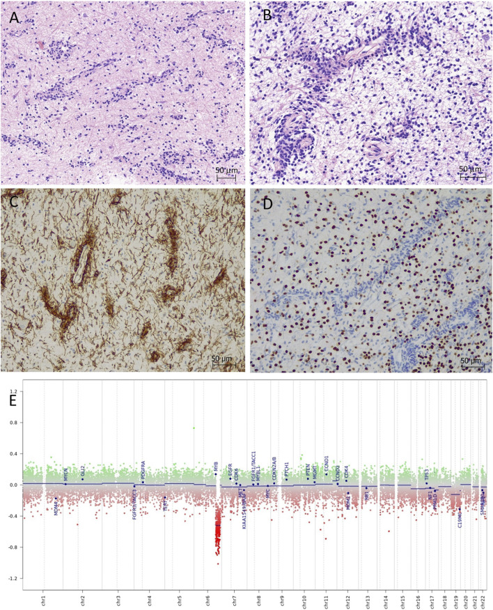
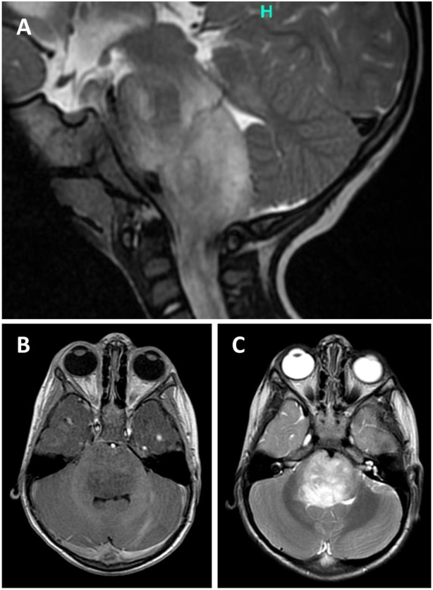
Introduction: Angiocentric gliomas (AG) in brainstem location are exceedingly rare and might cause differential diagnostic problems and uncertainty regarding the best therapeutic approach. Hereby, we describe the clinicopathological findings in a brainstem AG presenting in a toddler child and review the literature. Case report: A 2-year-old boy presented with 5 weeks history of gait disturbances, frequent falls, left-sided torticollis and swallowing problems. MRI head showed a T2-hyperintense, partly exophytic mass lesion centred in the pontomedullary region, raising the possibility of diffuse midline glioma. The exophytic component was partially resected by suboccipital craniotomy, leaving intact the infiltrative component. Ventriculoperitoneal shunt was implanted due to postoperative hydrocephalus. Histological examination revealed a moderately cellular tumour consisted of bland glial cells infiltrating the brain parenchyma and radially arranged around the blood vessels. By immunohistochemistry, the tumour strongly expressed S100 and GFAP in addition to intense nestin positivity, while OLIG2 was negative in the perivascular tumour cells. DNA methylation array profiled the tumour as "methylation class diffuse astrocytoma, MYB or MYBL1-altered subtype B (infratentorial)" and an in-frame MYB::QKI fusion was identified by RNA sequencing, confirming the diagnosis of angiocentric glioma. The patient has been initially treated with angiogenesis inhibitor and mTOR inhibitor, and now he is receiving palliative vinblastine. He is clinically stable on 9 months follow-up. Conclusion: Brainstem AG may cause a diagnostic problem, and the surgical and oncological management is challenging due to unresectability and lack of response to conventional chemo-radiation. In the future, genetically-tailored therapies might improve the prognosis.
期刊介绍:
Pathology & Oncology Research (POR) is an interdisciplinary Journal at the interface of pathology and oncology including the preclinical and translational research, diagnostics and therapy. Furthermore, POR is an international forum for the rapid communication of reviews, original research, critical and topical reports with excellence and novelty. Published quarterly, POR is dedicated to keeping scientists informed of developments on the selected biomedical fields bridging the gap between basic research and clinical medicine. It is a special aim for POR to promote pathological and oncological publishing activity of colleagues in the Central and East European region. The journal will be of interest to pathologists, and a broad range of experimental and clinical oncologists, and related experts. POR is supported by an acknowledged international advisory board and the Arányi Fundation for modern pathology.
 求助内容:
求助内容: 应助结果提醒方式:


