De novo expansion formation in the outer curvature of the internal carotid artery after flow diverter deployment for an infectious cavernous carotid aneurysm: illustrative case.
{"title":"De novo expansion formation in the outer curvature of the internal carotid artery after flow diverter deployment for an infectious cavernous carotid aneurysm: illustrative case.","authors":"Takuya Osuki, Hiroyuki Ikeda, Minami Uezato, Masanori Kinosada, Yoshitaka Kurosaki, Masaki Chin","doi":"10.3171/CASE23124","DOIUrl":null,"url":null,"abstract":"<p><strong>Background: </strong>Infectious aneurysms very rarely occur in the cavernous carotid artery. Recently, treatment by flow diverter implantation with preservation of the parent artery has been the treatment of choice.</p><p><strong>Observations: </strong>A 64-year-old woman presented with stenosis at the C5 segment of the left internal carotid artery (ICA), followed by ocular symptoms within 2 weeks, with a de novo aneurysm in the left cavernous carotid artery and wall irregularity with stenosis from the C2 to C5 segments of the left ICA. Antimicrobial therapy was given for 6 weeks, and a Pipeline Flex Shield was implanted. Angiography 6 months after treatment showed complete obliteration of the infectious aneurysm and improvement of the stenosis. However, de novo expansions were formed in the outer curvature of C3 and C4 segments of the ICA where the Pipeline device had been deployed.</p><p><strong>Lessons: </strong>Aneurysms that develop rapidly and show shape changes over time, accompanied by fever and inflammation, may be associated with an infection. Because of the fragility in the irregular wall of the parent vessel associated with infectious aneurysms, de novo expansion may form in the outer curvature of the parent vessel after flow diverter placement; thus, careful follow-up is necessary.</p>","PeriodicalId":16554,"journal":{"name":"Journal of Neurosurgery: Case Lessons","volume":"5 24","pages":""},"PeriodicalIF":0.0000,"publicationDate":"2023-06-12","publicationTypes":"Journal Article","fieldsOfStudy":null,"isOpenAccess":false,"openAccessPdf":"https://ftp.ncbi.nlm.nih.gov/pub/pmc/oa_pdf/c4/ee/CASE23124.PMC10550656.pdf","citationCount":"0","resultStr":null,"platform":"Semanticscholar","paperid":null,"PeriodicalName":"Journal of Neurosurgery: Case Lessons","FirstCategoryId":"1085","ListUrlMain":"https://doi.org/10.3171/CASE23124","RegionNum":0,"RegionCategory":null,"ArticlePicture":[],"TitleCN":null,"AbstractTextCN":null,"PMCID":null,"EPubDate":"","PubModel":"","JCR":"","JCRName":"","Score":null,"Total":0}
引用次数: 0
Abstract
Background: Infectious aneurysms very rarely occur in the cavernous carotid artery. Recently, treatment by flow diverter implantation with preservation of the parent artery has been the treatment of choice.
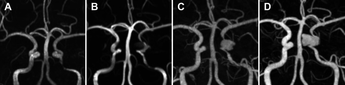
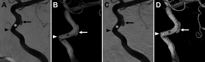
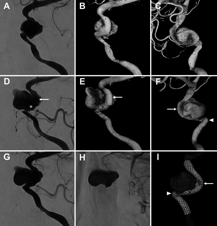
Observations: A 64-year-old woman presented with stenosis at the C5 segment of the left internal carotid artery (ICA), followed by ocular symptoms within 2 weeks, with a de novo aneurysm in the left cavernous carotid artery and wall irregularity with stenosis from the C2 to C5 segments of the left ICA. Antimicrobial therapy was given for 6 weeks, and a Pipeline Flex Shield was implanted. Angiography 6 months after treatment showed complete obliteration of the infectious aneurysm and improvement of the stenosis. However, de novo expansions were formed in the outer curvature of C3 and C4 segments of the ICA where the Pipeline device had been deployed.
Lessons: Aneurysms that develop rapidly and show shape changes over time, accompanied by fever and inflammation, may be associated with an infection. Because of the fragility in the irregular wall of the parent vessel associated with infectious aneurysms, de novo expansion may form in the outer curvature of the parent vessel after flow diverter placement; thus, careful follow-up is necessary.

 求助内容:
求助内容: 应助结果提醒方式:


