{"title":"Anatomical information of the lenticulostriate arteries on high-resolution 3D-TOF MRA at 3 T: comparison with 3D-DSA.","authors":"Sanae Takahashi, Miho Gomyo, Kazuhiro Tsuchiya, Tatsuya Yoshioka, Kuninori Kobayashi, Akihito Nakanishi, Kenichi Yokoyama","doi":"10.1007/s00276-023-03232-6","DOIUrl":null,"url":null,"abstract":"<p><strong>Purpose: </strong>As the lenticulostriate arteries (LSAs) perfuse neurologically important areas, it is necessary to accurately assess the origin and number of the LSAs before surgery. Although three-dimensional time-of-flight MR angiography (3D-TOF MRA) is a non-invasive procedure, it requires high-resolution (HR) images to depict the LSAs with a small diameter. Therefore, we performed 3D-TOF MRA with the maximum HR (HR-MRA) using a 3 T scanner to examine whether a good depiction of the LSAs, equivalent to that of digital subtraction angiography (DSA), could be obtained.</p><p><strong>Methods: </strong>Our study group comprised 16 consecutive patients who underwent HR-MRA and 3D-DSA. In both studies, we evaluated the localization of the origin from M1, M2, or A1 segments, their number of stems, and depiction.</p><p><strong>Results: </strong>There was no significant difference in the visualization of the LSAs between HR-MRA and 3D-DSA (P values; M1, M2, and A1 = 0.39, 0.69, and 0.69, respectively), and both the number of stems and the localization of the origin of the LSAs corresponded between the two examinations.</p><p><strong>Conclusion: </strong>HR-MRA at 3 T can depict the LSA well. It reveals the number of the LSA stems and the LSA origin comparatively with DSA.</p>","PeriodicalId":49296,"journal":{"name":"Surgical and Radiologic Anatomy","volume":" ","pages":"1287-1293"},"PeriodicalIF":1.2000,"publicationDate":"2023-10-01","publicationTypes":"Journal Article","fieldsOfStudy":null,"isOpenAccess":false,"openAccessPdf":"","citationCount":"0","resultStr":null,"platform":"Semanticscholar","paperid":null,"PeriodicalName":"Surgical and Radiologic Anatomy","FirstCategoryId":"3","ListUrlMain":"https://doi.org/10.1007/s00276-023-03232-6","RegionNum":4,"RegionCategory":"医学","ArticlePicture":[],"TitleCN":null,"AbstractTextCN":null,"PMCID":null,"EPubDate":"2023/8/24 0:00:00","PubModel":"Epub","JCR":"Q3","JCRName":"ANATOMY & MORPHOLOGY","Score":null,"Total":0}
引用次数: 0
Abstract
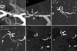
Purpose: As the lenticulostriate arteries (LSAs) perfuse neurologically important areas, it is necessary to accurately assess the origin and number of the LSAs before surgery. Although three-dimensional time-of-flight MR angiography (3D-TOF MRA) is a non-invasive procedure, it requires high-resolution (HR) images to depict the LSAs with a small diameter. Therefore, we performed 3D-TOF MRA with the maximum HR (HR-MRA) using a 3 T scanner to examine whether a good depiction of the LSAs, equivalent to that of digital subtraction angiography (DSA), could be obtained.
Methods: Our study group comprised 16 consecutive patients who underwent HR-MRA and 3D-DSA. In both studies, we evaluated the localization of the origin from M1, M2, or A1 segments, their number of stems, and depiction.
Results: There was no significant difference in the visualization of the LSAs between HR-MRA and 3D-DSA (P values; M1, M2, and A1 = 0.39, 0.69, and 0.69, respectively), and both the number of stems and the localization of the origin of the LSAs corresponded between the two examinations.
Conclusion: HR-MRA at 3 T can depict the LSA well. It reveals the number of the LSA stems and the LSA origin comparatively with DSA.
期刊介绍:
Anatomy is a morphological science which cannot fail to interest the clinician. The practical application of anatomical research to clinical problems necessitates special adaptation and selectivity in choosing from numerous international works. Although there is a tendency to believe that meaningful advances in anatomy are unlikely, constant revision is necessary. Surgical and Radiologic Anatomy, the first international journal of Clinical anatomy has been created in this spirit.
Its goal is to serve clinicians, regardless of speciality-physicians, surgeons, radiologists or other specialists-as an indispensable aid with which they can improve their knowledge of anatomy. Each issue includes: Original papers, review articles, articles on the anatomical bases of medical, surgical and radiological techniques, articles of normal radiologic anatomy, brief reviews of anatomical publications of clinical interest.
Particular attention is given to high quality illustrations, which are indispensable for a better understanding of anatomical problems.
Surgical and Radiologic Anatomy is a journal written by anatomists for clinicians with a special interest in anatomy.

 求助内容:
求助内容: 应助结果提醒方式:


