{"title":"Acute unilateral orbital varix thrombosis in preexisting bilateral orbital varices: illustrative case.","authors":"Chanokgarn Pichayawat, Weerawan Chokthaweesak, Sasikant Leelawongs, Ekachat Chanthanaphak, Oranan Tritanon, Bunyada Putthirangsiwong","doi":"10.3171/CASE23132","DOIUrl":null,"url":null,"abstract":"<p><strong>Objective: </strong>Orbital varix is a rare distensible orbital venous malformation. Most patients present with unilateral intermittent periorbital pain and positional proptosis that is exacerbated by the Valsalva maneuver. Complications include hemorrhage and thrombosis, leading to sudden painful proptosis and visual disturbance.</p><p><strong>Observations: </strong>A 42-year-old female with a history of bilateral intermittent painless proptosis that was accentuated by a postural head-down position presented with acute painful proptosis in her right eye. Ophthalmic examination revealed right eye proptosis with a bluish mass at the right upper eyelid, and another bluish mass at the left lower eyelid that was prominent during the Valsalva maneuver. Computed tomography scans of the orbits indicated right orbital varix thrombosis and left orbital varix. Surgical excision of the right thrombosed varix was performed along with intralesional bleomycin injection in the left orbital varix. Histopathological examination confirmed the right thrombosed varix diagnosis. The patient had good clinical improvement at the 6-month follow-up.</p><p><strong>Lessons: </strong>Orbital varices have a spectrum of clinical manifestations, from asymptomatic to severe visual loss. Most cases are successfully treated conservatively; however, in complicated cases, interventions with a multidisciplinary team approach such as sclerotherapy, embolization, and surgical excision should be considered.</p>","PeriodicalId":16554,"journal":{"name":"Journal of Neurosurgery: Case Lessons","volume":"5 25","pages":""},"PeriodicalIF":0.0000,"publicationDate":"2023-06-19","publicationTypes":"Journal Article","fieldsOfStudy":null,"isOpenAccess":false,"openAccessPdf":"https://ftp.ncbi.nlm.nih.gov/pub/pmc/oa_pdf/58/54/CASE23132.PMC10550533.pdf","citationCount":"0","resultStr":null,"platform":"Semanticscholar","paperid":null,"PeriodicalName":"Journal of Neurosurgery: Case Lessons","FirstCategoryId":"1085","ListUrlMain":"https://doi.org/10.3171/CASE23132","RegionNum":0,"RegionCategory":null,"ArticlePicture":[],"TitleCN":null,"AbstractTextCN":null,"PMCID":null,"EPubDate":"","PubModel":"","JCR":"","JCRName":"","Score":null,"Total":0}
引用次数: 0
Abstract
Objective: Orbital varix is a rare distensible orbital venous malformation. Most patients present with unilateral intermittent periorbital pain and positional proptosis that is exacerbated by the Valsalva maneuver. Complications include hemorrhage and thrombosis, leading to sudden painful proptosis and visual disturbance.
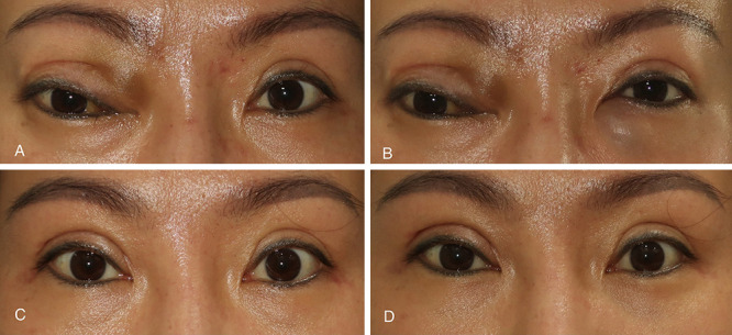
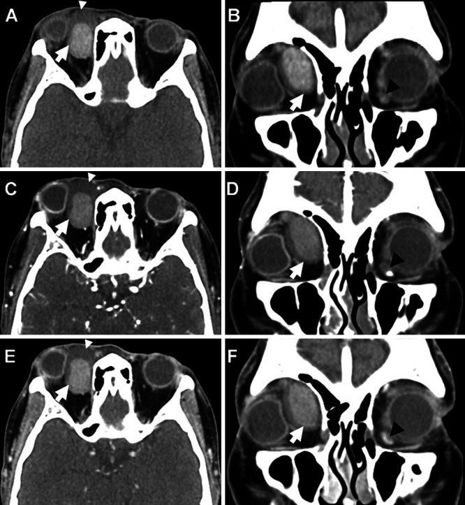
Observations: A 42-year-old female with a history of bilateral intermittent painless proptosis that was accentuated by a postural head-down position presented with acute painful proptosis in her right eye. Ophthalmic examination revealed right eye proptosis with a bluish mass at the right upper eyelid, and another bluish mass at the left lower eyelid that was prominent during the Valsalva maneuver. Computed tomography scans of the orbits indicated right orbital varix thrombosis and left orbital varix. Surgical excision of the right thrombosed varix was performed along with intralesional bleomycin injection in the left orbital varix. Histopathological examination confirmed the right thrombosed varix diagnosis. The patient had good clinical improvement at the 6-month follow-up.
Lessons: Orbital varices have a spectrum of clinical manifestations, from asymptomatic to severe visual loss. Most cases are successfully treated conservatively; however, in complicated cases, interventions with a multidisciplinary team approach such as sclerotherapy, embolization, and surgical excision should be considered.


 求助内容:
求助内容: 应助结果提醒方式:


