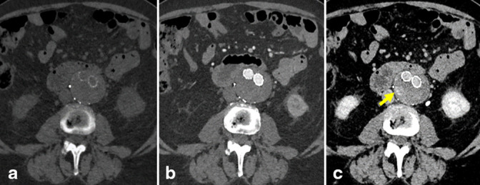
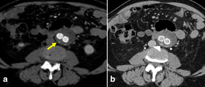
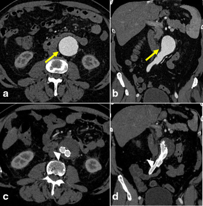
Surveillance imaging of type II endoleak with contrast-enhanced ultrasound.

IF 0.5 Q4 RADIOLOGY, NUCLEAR MEDICINE & MEDICAL IMAGING
Filip Jelenak, Jaspreet Hira, Rachita Khot
{"title":"Surveillance imaging of type II endoleak with contrast-enhanced ultrasound.","authors":"Filip Jelenak,&nbsp;Jaspreet Hira,&nbsp;Rachita Khot","doi":"10.1259/bjrcr.20230053","DOIUrl":null,"url":null,"abstract":"<p><p>Type II endoleak is the most common type of endoleak after endovascular repair of abdominal aortic aneurysm and has been reported in up to 20-50% of patients. Patients undergo lifelong surveillance of aortic graft stents to monitor for endoleak. Contrast-enhanced ultrasound can be an adjunct to CT angiography (CTA) which is the preferred imaging modality for surveillance. However, CT angiography introduces challenges of recurring cost, exposure to ionizing radiation, and the need for iodinated contrast dye. We report a case using CEUS for the detection of type II endoleak.</p>","PeriodicalId":45216,"journal":{"name":"BJR Case Reports","volume":null,"pages":null},"PeriodicalIF":0.5000,"publicationDate":"2023-08-01","publicationTypes":"Journal Article","fieldsOfStudy":null,"isOpenAccess":false,"openAccessPdf":"https://www.ncbi.nlm.nih.gov/pmc/articles/PMC10412912/pdf/","citationCount":"0","resultStr":null,"platform":"Semanticscholar","paperid":null,"PeriodicalName":"BJR Case Reports","FirstCategoryId":"1085","ListUrlMain":"https://doi.org/10.1259/bjrcr.20230053","RegionNum":0,"RegionCategory":null,"ArticlePicture":[],"TitleCN":null,"AbstractTextCN":null,"PMCID":null,"EPubDate":"","PubModel":"","JCR":"Q4","JCRName":"RADIOLOGY, NUCLEAR MEDICINE & MEDICAL IMAGING","Score":null,"Total":0}
引用次数: 0

Abstract

Type II endoleak is the most common type of endoleak after endovascular repair of abdominal aortic aneurysm and has been reported in up to 20-50% of patients. Patients undergo lifelong surveillance of aortic graft stents to monitor for endoleak. Contrast-enhanced ultrasound can be an adjunct to CT angiography (CTA) which is the preferred imaging modality for surveillance. However, CT angiography introduces challenges of recurring cost, exposure to ionizing radiation, and the need for iodinated contrast dye. We report a case using CEUS for the detection of type II endoleak.
Abstract Image

Abstract Image

Abstract Image

ⅱ型内漏的超声造影监测成像。
II型内漏是腹主动脉瘤血管内修复后最常见的内漏类型,据报道高达20-50%的患者发生了II型内漏。患者接受主动脉移植支架终身监测,以监测内漏。对比增强超声可以作为CT血管造影(CTA)的辅助手段,CTA是首选的监测成像方式。然而,CT血管造影带来了重复成本、暴露于电离辐射和需要碘化造影剂的挑战。我们报告一例使用超声造影检测II型内漏。
本文章由计算机程序翻译,如有差异,请以英文原文为准。
求助全文
约1分钟内获得全文 求助全文
来源期刊
BJR Case Reports
BJR Case Reports RADIOLOGY, NUCLEAR MEDICINE & MEDICAL IMAGING-
自引率
0.00%
发文量
77
审稿时长
11 weeks
×
引用
GB/T 7714-2015
复制
MLA
复制
APA
复制
导出至
BibTeX EndNote RefMan NoteFirst NoteExpress
×
提示
您的信息不完整,为了账户安全,请先补充。
现在去补充
×
提示
您因"违规操作"
具体请查看互助需知
我知道了
×
提示
确定
请完成安全验证×
copy
已复制链接
快去分享给好友吧!
我知道了
右上角分享
点击右上角分享
0
联系我们:info@booksci.cn Book学术提供免费学术资源搜索服务,方便国内外学者检索中英文文献。致力于提供最便捷和优质的服务体验。 Copyright © 2023 布克学术 All rights reserved.
京ICP备2023020795号-1
ghs 京公网安备 11010802042870号
Book学术文献互助
Book学术文献互助群
群 号:481959085
Book学术官方微信