Microneurosurgical treatment of a small perimesencephalic pure pial arterial malformation: an under-recognized etiology of angiographically occult subarachnoid hemorrhage. Illustrative case.
Robert C Sterner, Garret P Greeneway, Ufuk Erginoglu, Jaime L Martínez Santos, Mustafa K Baskaya
{"title":"Microneurosurgical treatment of a small perimesencephalic pure pial arterial malformation: an under-recognized etiology of angiographically occult subarachnoid hemorrhage. Illustrative case.","authors":"Robert C Sterner, Garret P Greeneway, Ufuk Erginoglu, Jaime L Martínez Santos, Mustafa K Baskaya","doi":"10.3171/CASE23246","DOIUrl":null,"url":null,"abstract":"<p><strong>Background: </strong>Pial arterial malformations (PAMs) are rare vascular lesions consisting of dilated tortuous arteries without venous drainage. Current PAM understanding is limited by the lesion's rarity, limited anatomopathological studies, and frequent misclassifications.</p><p><strong>Observations: </strong>A 23-year-old male experienced two spontaneous subarachnoid hemorrhages (SAHs) over 6 months with initially unremarkable diagnostic cerebral angiograms. Magnetic resonance imaging (MRI) and angiography after the second SAH revealed a small perimesencephalic ovoid lesion within the left crural cistern, between the left superior and posterior cerebral arteries, appearing to be an exophytic cavernoma, a thrombosed aneurysm, or a hemorrhagic tumor. Microsurgical resection was achieved with a pterional craniotomy and anterior clinoidectomy. The resected lesion was characteristic of a pure PAM arising from superior cerebellar arterial branches.</p><p><strong>Lessons: </strong>Small pure PAMs can be deceitfully dynamic lesions causing episodes of hemorrhage, complete thrombosis (angiographically occult), recanalization, and rehemorrhage. Small thrombosed vascular malformations or aneurysms should be included in differential diagnoses of angiographically occult SAH. MRI can be diagnostic, but the true angioarchitecture can only be elucidated with microneurosurgery. The only definitive cure is removal. The microneurosurgical strategy should account for worst-case scenarios, provide adequate skull base exposures, and include bypass revascularization options when thrombosed aneurysms are encountered.</p>","PeriodicalId":16554,"journal":{"name":"Journal of Neurosurgery: Case Lessons","volume":"6 5","pages":""},"PeriodicalIF":0.0000,"publicationDate":"2023-07-31","publicationTypes":"Journal Article","fieldsOfStudy":null,"isOpenAccess":false,"openAccessPdf":"https://ftp.ncbi.nlm.nih.gov/pub/pmc/oa_pdf/81/a8/CASE23246.PMC10555581.pdf","citationCount":"0","resultStr":null,"platform":"Semanticscholar","paperid":null,"PeriodicalName":"Journal of Neurosurgery: Case Lessons","FirstCategoryId":"1085","ListUrlMain":"https://doi.org/10.3171/CASE23246","RegionNum":0,"RegionCategory":null,"ArticlePicture":[],"TitleCN":null,"AbstractTextCN":null,"PMCID":null,"EPubDate":"","PubModel":"","JCR":"","JCRName":"","Score":null,"Total":0}
引用次数: 0
Abstract
Background: Pial arterial malformations (PAMs) are rare vascular lesions consisting of dilated tortuous arteries without venous drainage. Current PAM understanding is limited by the lesion's rarity, limited anatomopathological studies, and frequent misclassifications.
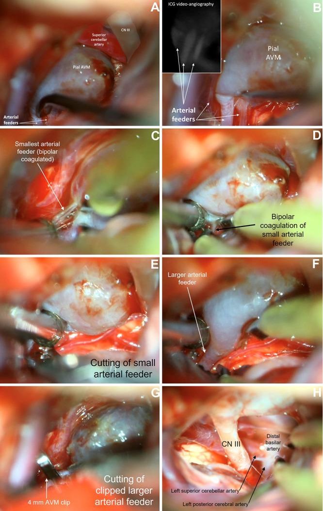
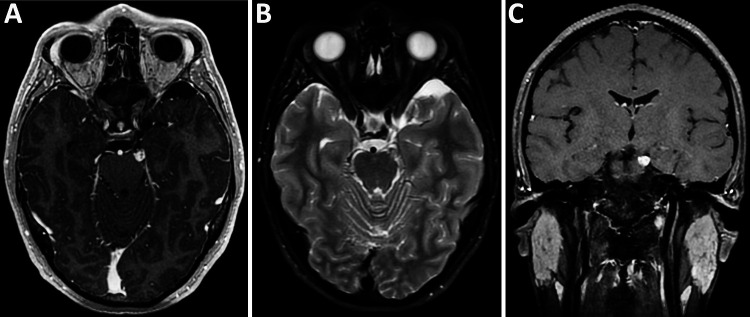
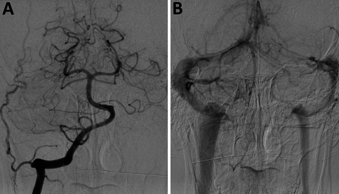
Observations: A 23-year-old male experienced two spontaneous subarachnoid hemorrhages (SAHs) over 6 months with initially unremarkable diagnostic cerebral angiograms. Magnetic resonance imaging (MRI) and angiography after the second SAH revealed a small perimesencephalic ovoid lesion within the left crural cistern, between the left superior and posterior cerebral arteries, appearing to be an exophytic cavernoma, a thrombosed aneurysm, or a hemorrhagic tumor. Microsurgical resection was achieved with a pterional craniotomy and anterior clinoidectomy. The resected lesion was characteristic of a pure PAM arising from superior cerebellar arterial branches.
Lessons: Small pure PAMs can be deceitfully dynamic lesions causing episodes of hemorrhage, complete thrombosis (angiographically occult), recanalization, and rehemorrhage. Small thrombosed vascular malformations or aneurysms should be included in differential diagnoses of angiographically occult SAH. MRI can be diagnostic, but the true angioarchitecture can only be elucidated with microneurosurgery. The only definitive cure is removal. The microneurosurgical strategy should account for worst-case scenarios, provide adequate skull base exposures, and include bypass revascularization options when thrombosed aneurysms are encountered.

 求助内容:
求助内容: 应助结果提醒方式:


