Nahom Teferi, Ajmain Chowdhury, Sarah Lee, Meron Challa, Lukasz Weiner, Sarah Auerbach, Mahil Rao, Brian J Dlouhy
{"title":"Pediatric brainstem abscess successfully treated with stereotactic aspiration: illustrative case.","authors":"Nahom Teferi, Ajmain Chowdhury, Sarah Lee, Meron Challa, Lukasz Weiner, Sarah Auerbach, Mahil Rao, Brian J Dlouhy","doi":"10.3171/CASE23262","DOIUrl":null,"url":null,"abstract":"<p><strong>Background: </strong>Pediatric brainstem abscesses are rare entities that account for 1% of all brain abscesses and, when diagnosed, constitute a neurosurgical emergency.</p><p><strong>Observations: </strong>A previously healthy 11-year-old male presented with several days of worsening headache, confusion, and ataxia. Brain magnetic resonance imaging (MRI) revealed a midbrain and pons lesion. The patient subsequently had a rapid neurological decline with loss of consciousness and brainstem function. Follow-up MRI revealed significant enlargement of the brainstem lesion with extension into the pons, midbrain, and thalamus, with greater concerns for an abscess rather than a tumor or an inflammatory process. He was taken for an emergent stereotactic aspiration of the abscess, and broad-spectrum antibiotics were initiated. He had neurological improvement, which subsequently declined 5 days later with brain MRI revealing an increase in the brainstem abscess, which required a second stereotactic aspiration. After rehabilitation, he made a significant neurological recovery.</p><p><strong>Lessons: </strong>Pediatric brainstem abscesses are rare pathologies, and a high index of suspicion is needed in patients presenting with a brainstem lesion mimicking tumor but with rapid neurological decline despite no other evidence of infection or infectious/inflammatory markers. Stereotactic aspiration is required for large lesions to target the antibiotic treatment and as an adjunct to broad-spectrum antibiotics.</p>","PeriodicalId":16554,"journal":{"name":"Journal of Neurosurgery: Case Lessons","volume":"6 6","pages":""},"PeriodicalIF":0.0000,"publicationDate":"2023-08-07","publicationTypes":"Journal Article","fieldsOfStudy":null,"isOpenAccess":false,"openAccessPdf":"https://ftp.ncbi.nlm.nih.gov/pub/pmc/oa_pdf/24/a8/CASE23262.PMC10555595.pdf","citationCount":"0","resultStr":null,"platform":"Semanticscholar","paperid":null,"PeriodicalName":"Journal of Neurosurgery: Case Lessons","FirstCategoryId":"1085","ListUrlMain":"https://doi.org/10.3171/CASE23262","RegionNum":0,"RegionCategory":null,"ArticlePicture":[],"TitleCN":null,"AbstractTextCN":null,"PMCID":null,"EPubDate":"","PubModel":"","JCR":"","JCRName":"","Score":null,"Total":0}
引用次数: 0
Abstract
Background: Pediatric brainstem abscesses are rare entities that account for 1% of all brain abscesses and, when diagnosed, constitute a neurosurgical emergency.
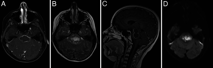
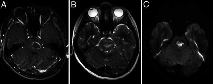
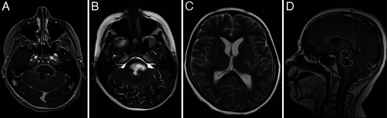
Observations: A previously healthy 11-year-old male presented with several days of worsening headache, confusion, and ataxia. Brain magnetic resonance imaging (MRI) revealed a midbrain and pons lesion. The patient subsequently had a rapid neurological decline with loss of consciousness and brainstem function. Follow-up MRI revealed significant enlargement of the brainstem lesion with extension into the pons, midbrain, and thalamus, with greater concerns for an abscess rather than a tumor or an inflammatory process. He was taken for an emergent stereotactic aspiration of the abscess, and broad-spectrum antibiotics were initiated. He had neurological improvement, which subsequently declined 5 days later with brain MRI revealing an increase in the brainstem abscess, which required a second stereotactic aspiration. After rehabilitation, he made a significant neurological recovery.
Lessons: Pediatric brainstem abscesses are rare pathologies, and a high index of suspicion is needed in patients presenting with a brainstem lesion mimicking tumor but with rapid neurological decline despite no other evidence of infection or infectious/inflammatory markers. Stereotactic aspiration is required for large lesions to target the antibiotic treatment and as an adjunct to broad-spectrum antibiotics.

 求助内容:
求助内容: 应助结果提醒方式:


