{"title":"光学相干断层扫描血管造影对青少年单纯性近视和非单纯性近视视网膜微血管密度的比较。","authors":"Kemal Bayrakçeken","doi":"10.5152/eurasianjmed.2023.22278","DOIUrl":null,"url":null,"abstract":"<p><strong>Objective: </strong>The aim of this study is to investigate whether there is a difference in retinal microvascularization between adolescents with and without simple myopia using optical coherence tomography angiography.</p><p><strong>Materials and methods: </strong>Thirty-four eyes of 34 patients aged 12-18 years diagnosed with school-age simple myopia (0-6 diopters), and 34 eyes of 34 healthy controls of similar ages were included in this retrospective study. The ocular, optical coherence tomography, and optical coherence tomography angiography findings of the participants were recorded.</p><p><strong>Results: </strong>The simple myopia group had statistically thicker inferior ganglion cell complex thicknesses compared to the controls (P =.038). The macular map values did not statistically significantly differ between the 2 groups. The foveal avascular zone area (P =.038) and circularity index (P =.022) values were statistically lower in the simple myopia group compared to the control group. The superficial capillary plexus outer and inner ring vessel density (%) (superior and nasal) showed statistically significant differences (outer ring superior/ nasal P=.004/P = .037; inner ring superior/nasal P =.014/P= .046).</p><p><strong>Conclusion: </strong>Similar to high myopia, vascular density in the macula decreases as the axial length and spherical equivalent increase in simple myopia.</p>","PeriodicalId":53592,"journal":{"name":"Eurasian Journal of Medicine","volume":null,"pages":null},"PeriodicalIF":0.9000,"publicationDate":"2023-02-01","publicationTypes":"Journal Article","fieldsOfStudy":null,"isOpenAccess":false,"openAccessPdf":"https://www.ncbi.nlm.nih.gov/pmc/articles/PMC10081140/pdf/","citationCount":"1","resultStr":"{\"title\":\"Comparison of Retinal Microvascular Vascular Density Between Adolescents With and Without Simple Myopia Using Optical Coherence Tomography Angiography.\",\"authors\":\"Kemal Bayrakçeken\",\"doi\":\"10.5152/eurasianjmed.2023.22278\",\"DOIUrl\":null,\"url\":null,\"abstract\":\"<p><strong>Objective: </strong>The aim of this study is to investigate whether there is a difference in retinal microvascularization between adolescents with and without simple myopia using optical coherence tomography angiography.</p><p><strong>Materials and methods: </strong>Thirty-four eyes of 34 patients aged 12-18 years diagnosed with school-age simple myopia (0-6 diopters), and 34 eyes of 34 healthy controls of similar ages were included in this retrospective study. The ocular, optical coherence tomography, and optical coherence tomography angiography findings of the participants were recorded.</p><p><strong>Results: </strong>The simple myopia group had statistically thicker inferior ganglion cell complex thicknesses compared to the controls (P =.038). The macular map values did not statistically significantly differ between the 2 groups. The foveal avascular zone area (P =.038) and circularity index (P =.022) values were statistically lower in the simple myopia group compared to the control group. The superficial capillary plexus outer and inner ring vessel density (%) (superior and nasal) showed statistically significant differences (outer ring superior/ nasal P=.004/P = .037; inner ring superior/nasal P =.014/P= .046).</p><p><strong>Conclusion: </strong>Similar to high myopia, vascular density in the macula decreases as the axial length and spherical equivalent increase in simple myopia.</p>\",\"PeriodicalId\":53592,\"journal\":{\"name\":\"Eurasian Journal of Medicine\",\"volume\":null,\"pages\":null},\"PeriodicalIF\":0.9000,\"publicationDate\":\"2023-02-01\",\"publicationTypes\":\"Journal Article\",\"fieldsOfStudy\":null,\"isOpenAccess\":false,\"openAccessPdf\":\"https://www.ncbi.nlm.nih.gov/pmc/articles/PMC10081140/pdf/\",\"citationCount\":\"1\",\"resultStr\":null,\"platform\":\"Semanticscholar\",\"paperid\":null,\"PeriodicalName\":\"Eurasian Journal of Medicine\",\"FirstCategoryId\":\"1085\",\"ListUrlMain\":\"https://doi.org/10.5152/eurasianjmed.2023.22278\",\"RegionNum\":0,\"RegionCategory\":null,\"ArticlePicture\":[],\"TitleCN\":null,\"AbstractTextCN\":null,\"PMCID\":null,\"EPubDate\":\"\",\"PubModel\":\"\",\"JCR\":\"Q3\",\"JCRName\":\"MEDICINE, GENERAL & INTERNAL\",\"Score\":null,\"Total\":0}","platform":"Semanticscholar","paperid":null,"PeriodicalName":"Eurasian Journal of Medicine","FirstCategoryId":"1085","ListUrlMain":"https://doi.org/10.5152/eurasianjmed.2023.22278","RegionNum":0,"RegionCategory":null,"ArticlePicture":[],"TitleCN":null,"AbstractTextCN":null,"PMCID":null,"EPubDate":"","PubModel":"","JCR":"Q3","JCRName":"MEDICINE, GENERAL & INTERNAL","Score":null,"Total":0}
引用次数: 1
摘要
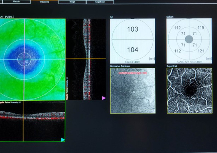
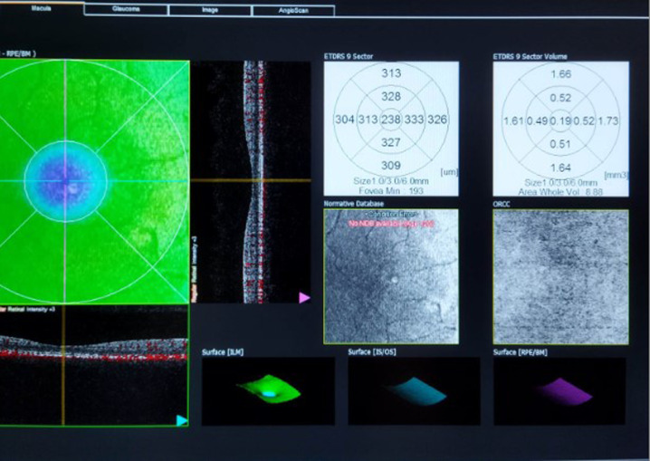
目的:本研究的目的是通过光学相干断层扫描血管成像研究青少年单纯性近视和非单纯性近视的视网膜微血管形成是否存在差异。材料与方法:回顾性研究34例12-18岁学龄期单纯性近视(0-6屈光度)患者34只眼和34例年龄相近的健康对照者34只眼。记录参与者的眼部、光学相干断层扫描和光学相干断层扫描血管造影结果。结果:单纯性近视组下神经节细胞复合体厚度较对照组有统计学意义(P = 0.038)。两组间黄斑图值差异无统计学意义。单纯性近视组中央凹无血管区面积(P = 0.038)和圆度指数(P = 0.022)值均低于对照组,差异有统计学意义。浅表毛细血管丛外、内环血管密度(%)(上、鼻)差异有统计学意义(外、上、鼻P=。004/ p = 0.037;内环上/鼻P =。014 / P = .046)。结论:与高度近视相似,单纯性近视黄斑血管密度随着眼轴长度和球当量的增加而降低。
Comparison of Retinal Microvascular Vascular Density Between Adolescents With and Without Simple Myopia Using Optical Coherence Tomography Angiography.
Objective: The aim of this study is to investigate whether there is a difference in retinal microvascularization between adolescents with and without simple myopia using optical coherence tomography angiography.
Materials and methods: Thirty-four eyes of 34 patients aged 12-18 years diagnosed with school-age simple myopia (0-6 diopters), and 34 eyes of 34 healthy controls of similar ages were included in this retrospective study. The ocular, optical coherence tomography, and optical coherence tomography angiography findings of the participants were recorded.
Results: The simple myopia group had statistically thicker inferior ganglion cell complex thicknesses compared to the controls (P =.038). The macular map values did not statistically significantly differ between the 2 groups. The foveal avascular zone area (P =.038) and circularity index (P =.022) values were statistically lower in the simple myopia group compared to the control group. The superficial capillary plexus outer and inner ring vessel density (%) (superior and nasal) showed statistically significant differences (outer ring superior/ nasal P=.004/P = .037; inner ring superior/nasal P =.014/P= .046).
Conclusion: Similar to high myopia, vascular density in the macula decreases as the axial length and spherical equivalent increase in simple myopia.
期刊介绍:
Eurasian Journal of Medicine (Eurasian J Med) is an international, scientific, open access periodical published by independent, unbiased, and triple-blinded peer-review principles. The journal is the official publication of Atatürk University School of Medicine and published triannually in February, June, and October. The publication language of the journal is English. The aim of the Eurasian Journal of Medicine is to publish original research papers of the highest scientific and clinical value in all medical fields. The Eurasian J Med also includes reviews, editorial short notes and letters to the editor that either as a comment related to recently published articles in our journal or as a case report. The target audience of the journal includes researchers, physicians and healthcare professionals who are interested or working in in all medical disciplines.


 求助内容:
求助内容: 应助结果提醒方式:


