Amit Maheshwari, Dhruv Nilesh Chawda, Ashish Kushwah, Rajesh Kumar Agarwal, Amesh Kr Golwara, Prateek Bhushan Dixit
{"title":"骨根外和骨根间微型种植体下颌弓远端移位和应力分布模式的比较评估:三维有限元研究。","authors":"Amit Maheshwari, Dhruv Nilesh Chawda, Ashish Kushwah, Rajesh Kumar Agarwal, Amesh Kr Golwara, Prateek Bhushan Dixit","doi":"10.1590/2177-6709.28.2.e2321373.oar","DOIUrl":null,"url":null,"abstract":"<p><strong>Objective: </strong>To compare the initial stress distribution and displacement on mandibular dentition using extra and inter-radicular mini-implants for arch distalization, by means of finite element analysis.</p><p><strong>Methods: </strong>For this study, two finite element models of the mandible were designed. The models consisted of periodontal ligament (PDL) and alveolar bone of all teeth until second molars. In the Case 1, bilateral extra-radicular buccal-shelf stainless steel mini-implants (10.0-mm length; 2.0-mm diameter) were placed between first and second permanent molars. In the Case 2, bilateral inter-radicular stainless steel mini-implants (10.0-mm length; 1.5-mm diameter) were placed between second premolar and first permanent molar. Power hook was attached between canine and first premolar at a fixed height of 8mm. In the two cases, 200g of distalization force was applied. ANSYS v. 12.1 software was used to analyze and compare von Mises stress and displacement in the mandibular dentition, PDL and bone.</p><p><strong>Results: </strong>Higher stresses were observed in mandibular dentition with the inter-radicular implant system. The amount of von Mises stress was higher for cortical bone (85.66MPa) and cancellous bone (3.64MPa) in Case 2, in comparison to cortical bone (41.93MPa) and cancellous bone (3.43MPa) in Case 1. The amount of arch distalization was higher for mandible in Case 1 (0.028mm), in comparison to Case 2 (0.026mm).</p><p><strong>Conclusion: </strong>Both systems were clinically safe, but extra-radicular implants showed more effective and controlled distalization pattern, in comparison to inter-radicular implants, in Class III malocclusion treatment.</p>","PeriodicalId":38720,"journal":{"name":"Dental Press Journal of Orthodontics","volume":"28 2","pages":"e2321373"},"PeriodicalIF":0.0000,"publicationDate":"2023-01-01","publicationTypes":"Journal Article","fieldsOfStudy":null,"isOpenAccess":false,"openAccessPdf":"https://www.ncbi.nlm.nih.gov/pmc/articles/PMC10229115/pdf/","citationCount":"1","resultStr":"{\"title\":\"Comparative evaluation of displacement and stress distribution pattern during mandibular arch distalization with extra and inter-radicular mini-implants: a three-dimensional finite element study.\",\"authors\":\"Amit Maheshwari, Dhruv Nilesh Chawda, Ashish Kushwah, Rajesh Kumar Agarwal, Amesh Kr Golwara, Prateek Bhushan Dixit\",\"doi\":\"10.1590/2177-6709.28.2.e2321373.oar\",\"DOIUrl\":null,\"url\":null,\"abstract\":\"<p><strong>Objective: </strong>To compare the initial stress distribution and displacement on mandibular dentition using extra and inter-radicular mini-implants for arch distalization, by means of finite element analysis.</p><p><strong>Methods: </strong>For this study, two finite element models of the mandible were designed. The models consisted of periodontal ligament (PDL) and alveolar bone of all teeth until second molars. In the Case 1, bilateral extra-radicular buccal-shelf stainless steel mini-implants (10.0-mm length; 2.0-mm diameter) were placed between first and second permanent molars. In the Case 2, bilateral inter-radicular stainless steel mini-implants (10.0-mm length; 1.5-mm diameter) were placed between second premolar and first permanent molar. Power hook was attached between canine and first premolar at a fixed height of 8mm. In the two cases, 200g of distalization force was applied. ANSYS v. 12.1 software was used to analyze and compare von Mises stress and displacement in the mandibular dentition, PDL and bone.</p><p><strong>Results: </strong>Higher stresses were observed in mandibular dentition with the inter-radicular implant system. The amount of von Mises stress was higher for cortical bone (85.66MPa) and cancellous bone (3.64MPa) in Case 2, in comparison to cortical bone (41.93MPa) and cancellous bone (3.43MPa) in Case 1. The amount of arch distalization was higher for mandible in Case 1 (0.028mm), in comparison to Case 2 (0.026mm).</p><p><strong>Conclusion: </strong>Both systems were clinically safe, but extra-radicular implants showed more effective and controlled distalization pattern, in comparison to inter-radicular implants, in Class III malocclusion treatment.</p>\",\"PeriodicalId\":38720,\"journal\":{\"name\":\"Dental Press Journal of Orthodontics\",\"volume\":\"28 2\",\"pages\":\"e2321373\"},\"PeriodicalIF\":0.0000,\"publicationDate\":\"2023-01-01\",\"publicationTypes\":\"Journal Article\",\"fieldsOfStudy\":null,\"isOpenAccess\":false,\"openAccessPdf\":\"https://www.ncbi.nlm.nih.gov/pmc/articles/PMC10229115/pdf/\",\"citationCount\":\"1\",\"resultStr\":null,\"platform\":\"Semanticscholar\",\"paperid\":null,\"PeriodicalName\":\"Dental Press Journal of Orthodontics\",\"FirstCategoryId\":\"1085\",\"ListUrlMain\":\"https://doi.org/10.1590/2177-6709.28.2.e2321373.oar\",\"RegionNum\":0,\"RegionCategory\":null,\"ArticlePicture\":[],\"TitleCN\":null,\"AbstractTextCN\":null,\"PMCID\":null,\"EPubDate\":\"\",\"PubModel\":\"\",\"JCR\":\"Q2\",\"JCRName\":\"Medicine\",\"Score\":null,\"Total\":0}","platform":"Semanticscholar","paperid":null,"PeriodicalName":"Dental Press Journal of Orthodontics","FirstCategoryId":"1085","ListUrlMain":"https://doi.org/10.1590/2177-6709.28.2.e2321373.oar","RegionNum":0,"RegionCategory":null,"ArticlePicture":[],"TitleCN":null,"AbstractTextCN":null,"PMCID":null,"EPubDate":"","PubModel":"","JCR":"Q2","JCRName":"Medicine","Score":null,"Total":0}
引用次数: 1
摘要
目的:通过有限元分析比较根外与根内微型种植体用于下颌牙列弓远端的初始应力分布和位移。方法:设计2个下颌骨有限元模型。模型包括所有牙齿的牙周韧带(PDL)和牙槽骨,直到第二磨牙。在病例1中,双侧根外颊架不锈钢微型种植体(10.0 mm长度;直径2.0 mm)放置在第一恒磨牙和第二恒磨牙之间。在病例2中,双侧根间不锈钢微型种植体(10.0 mm长度;直径1.5 mm)放置在第二前磨牙和第一恒磨牙之间。动力钩固定在犬齿与第一前磨牙之间,固定高度为8mm。在这两种情况下,施加了200g的远端力。采用ANSYS v. 12.1软件分析比较下颌牙列、PDL和骨的von Mises应力和位移。结果:根间种植系统对下颌牙列有较高的应力。病例2中皮质骨(85.66MPa)和松质骨(3.64MPa)的von Mises应力量高于病例1中皮质骨(41.93MPa)和松质骨(3.43MPa)。病例1的下颌弓远距量(0.028mm)高于病例2 (0.026mm)。结论:两种系统在临床上都是安全的,但根外种植体与根内种植体相比,在治疗III类错颌时表现出更有效和可控的远端模式。
Comparative evaluation of displacement and stress distribution pattern during mandibular arch distalization with extra and inter-radicular mini-implants: a three-dimensional finite element study.
Objective: To compare the initial stress distribution and displacement on mandibular dentition using extra and inter-radicular mini-implants for arch distalization, by means of finite element analysis.
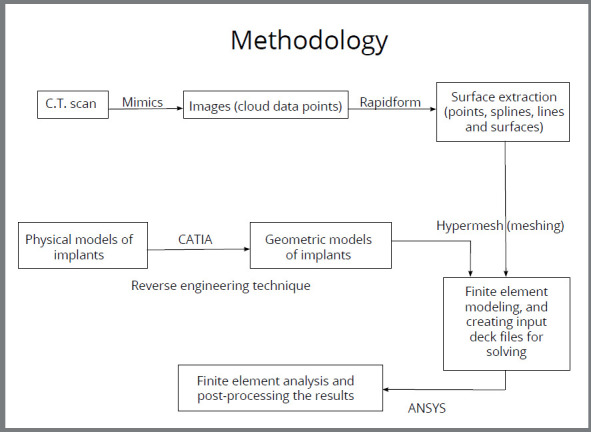
Methods: For this study, two finite element models of the mandible were designed. The models consisted of periodontal ligament (PDL) and alveolar bone of all teeth until second molars. In the Case 1, bilateral extra-radicular buccal-shelf stainless steel mini-implants (10.0-mm length; 2.0-mm diameter) were placed between first and second permanent molars. In the Case 2, bilateral inter-radicular stainless steel mini-implants (10.0-mm length; 1.5-mm diameter) were placed between second premolar and first permanent molar. Power hook was attached between canine and first premolar at a fixed height of 8mm. In the two cases, 200g of distalization force was applied. ANSYS v. 12.1 software was used to analyze and compare von Mises stress and displacement in the mandibular dentition, PDL and bone.
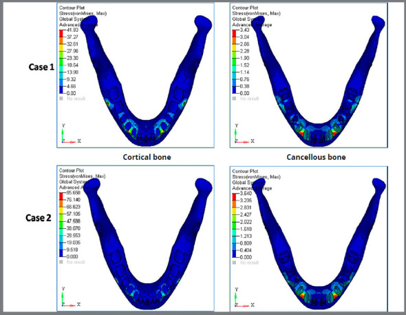
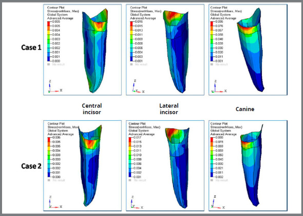
Results: Higher stresses were observed in mandibular dentition with the inter-radicular implant system. The amount of von Mises stress was higher for cortical bone (85.66MPa) and cancellous bone (3.64MPa) in Case 2, in comparison to cortical bone (41.93MPa) and cancellous bone (3.43MPa) in Case 1. The amount of arch distalization was higher for mandible in Case 1 (0.028mm), in comparison to Case 2 (0.026mm).
Conclusion: Both systems were clinically safe, but extra-radicular implants showed more effective and controlled distalization pattern, in comparison to inter-radicular implants, in Class III malocclusion treatment.
期刊介绍:
The Dental Press Journal of Orthodontics publishes scientific research articles, significant reviews, clinical and technical case reports, brief communications, and other materials related to Orthodontics and Facial Orthopedics.

 求助内容:
求助内容: 应助结果提醒方式:


