基于改良Sca/eS的小鼠耳蜗三维共聚焦显微镜成像

IF 1.2 4区 医学 Q3 ANATOMY & MORPHOLOGY
Shinji Urata, Shigeo Okabe
{"title":"基于改良Sca/eS的小鼠耳蜗三维共聚焦显微镜成像","authors":"Shinji Urata,&nbsp;Shigeo Okabe","doi":"10.1007/s12565-023-00703-z","DOIUrl":null,"url":null,"abstract":"<div><p>The three-dimensional stria vascularis (SV) and cochlear blood vessel structure is essential for inner ear function. Here, modified Sca/eS, a sorbitol-based optical-clearing method, was reported to visualize SV and vascular structure in the intact mouse cochlea. Cochlear macrophages as well as perivascular-resident macrophage-like melanocytes were detected as GFP-positive cells of the CX3CR1<sup>+/GFP</sup> mice. This study’s method was effective in elucidating inner ear function under both physiological and pathological conditions.</p></div>","PeriodicalId":7816,"journal":{"name":"Anatomical Science International","volume":null,"pages":null},"PeriodicalIF":1.2000,"publicationDate":"2023-02-11","publicationTypes":"Journal Article","fieldsOfStudy":null,"isOpenAccess":false,"openAccessPdf":"https://link.springer.com/content/pdf/10.1007/s12565-023-00703-z.pdf","citationCount":"0","resultStr":"{\"title\":\"Three-dimensional mouse cochlea imaging based on the modified Sca/eS using confocal microscopy\",\"authors\":\"Shinji Urata,&nbsp;Shigeo Okabe\",\"doi\":\"10.1007/s12565-023-00703-z\",\"DOIUrl\":null,\"url\":null,\"abstract\":\"<div><p>The three-dimensional stria vascularis (SV) and cochlear blood vessel structure is essential for inner ear function. Here, modified Sca/eS, a sorbitol-based optical-clearing method, was reported to visualize SV and vascular structure in the intact mouse cochlea. Cochlear macrophages as well as perivascular-resident macrophage-like melanocytes were detected as GFP-positive cells of the CX3CR1<sup>+/GFP</sup> mice. This study’s method was effective in elucidating inner ear function under both physiological and pathological conditions.</p></div>\",\"PeriodicalId\":7816,\"journal\":{\"name\":\"Anatomical Science International\",\"volume\":null,\"pages\":null},\"PeriodicalIF\":1.2000,\"publicationDate\":\"2023-02-11\",\"publicationTypes\":\"Journal Article\",\"fieldsOfStudy\":null,\"isOpenAccess\":false,\"openAccessPdf\":\"https://link.springer.com/content/pdf/10.1007/s12565-023-00703-z.pdf\",\"citationCount\":\"0\",\"resultStr\":null,\"platform\":\"Semanticscholar\",\"paperid\":null,\"PeriodicalName\":\"Anatomical Science International\",\"FirstCategoryId\":\"3\",\"ListUrlMain\":\"https://link.springer.com/article/10.1007/s12565-023-00703-z\",\"RegionNum\":4,\"RegionCategory\":\"医学\",\"ArticlePicture\":[],\"TitleCN\":null,\"AbstractTextCN\":null,\"PMCID\":null,\"EPubDate\":\"\",\"PubModel\":\"\",\"JCR\":\"Q3\",\"JCRName\":\"ANATOMY & MORPHOLOGY\",\"Score\":null,\"Total\":0}","platform":"Semanticscholar","paperid":null,"PeriodicalName":"Anatomical Science International","FirstCategoryId":"3","ListUrlMain":"https://link.springer.com/article/10.1007/s12565-023-00703-z","RegionNum":4,"RegionCategory":"医学","ArticlePicture":[],"TitleCN":null,"AbstractTextCN":null,"PMCID":null,"EPubDate":"","PubModel":"","JCR":"Q3","JCRName":"ANATOMY & MORPHOLOGY","Score":null,"Total":0}
引用次数: 0

摘要

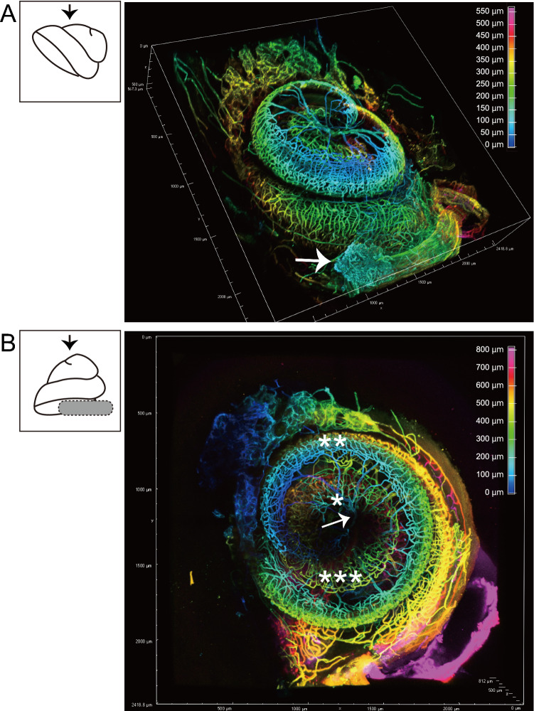
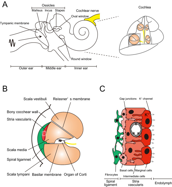
三维血管纹(SV)和耳蜗血管结构对内耳功能至关重要。本文报道了一种改良的Sca/eS,一种基于山梨醇的光学清除方法,用于显示完整小鼠耳蜗中的SV和血管结构。耳蜗巨噬细胞以及血管周围驻留的巨噬细胞样黑素细胞被检测为CX3CR1+/GFP小鼠的GFP阳性细胞。本研究的方法在生理和病理条件下都能有效地阐明内耳功能。
本文章由计算机程序翻译,如有差异,请以英文原文为准。

Three-dimensional mouse cochlea imaging based on the modified Sca/eS using confocal microscopy

Three-dimensional mouse cochlea imaging based on the modified Sca/eS using confocal microscopy

Three-dimensional mouse cochlea imaging based on the modified Sca/eS using confocal microscopy

Three-dimensional mouse cochlea imaging based on the modified Sca/eS using confocal microscopy

The three-dimensional stria vascularis (SV) and cochlear blood vessel structure is essential for inner ear function. Here, modified Sca/eS, a sorbitol-based optical-clearing method, was reported to visualize SV and vascular structure in the intact mouse cochlea. Cochlear macrophages as well as perivascular-resident macrophage-like melanocytes were detected as GFP-positive cells of the CX3CR1+/GFP mice. This study’s method was effective in elucidating inner ear function under both physiological and pathological conditions.

求助全文
通过发布文献求助,成功后即可免费获取论文全文。 去求助
来源期刊
Anatomical Science International
Anatomical Science International 医学-解剖学与形态学
CiteScore
2.80
自引率
8.30%
发文量
50
审稿时长
>12 weeks
期刊介绍: The official English journal of the Japanese Association of Anatomists, Anatomical Science International (formerly titled Kaibogaku Zasshi) publishes original research articles dealing with morphological sciences. Coverage in the journal includes molecular, cellular, histological and gross anatomical studies on humans and on normal and experimental animals, as well as functional morphological, biochemical, physiological and behavioral studies if they include morphological analysis.
×
引用
GB/T 7714-2015
复制
MLA
复制
APA
复制
导出至
BibTeX EndNote RefMan NoteFirst NoteExpress
×
提示
您的信息不完整,为了账户安全,请先补充。
现在去补充
×
提示
您因"违规操作"
具体请查看互助需知
我知道了
×
提示
确定
请完成安全验证×
copy
已复制链接
快去分享给好友吧!
我知道了
右上角分享
点击右上角分享
0
联系我们:info@booksci.cn Book学术提供免费学术资源搜索服务,方便国内外学者检索中英文文献。致力于提供最便捷和优质的服务体验。 Copyright © 2023 布克学术 All rights reserved.
京ICP备2023020795号-1
ghs 京公网安备 11010802042870号
Book学术文献互助
Book学术文献互助群
群 号:481959085
Book学术官方微信