{"title":"一种新的宽范围连续切片和三维重建相结合的方法,该方法同时显示了宏观层面的动力学和微观层面的相互作用,试图分析女性盆底","authors":"Satoru Muro, Keiichi Akita","doi":"10.1007/s12565-023-00710-0","DOIUrl":null,"url":null,"abstract":"<div><p>The present report presents details of the method for combining wide-range serial sectioning and 3D reconstruction using an adult cadaver. For several decades, anatomists have utilized a variety of non-destructive three-dimensional (3D) visualization methods to complement gross anatomical analysis methods. These include vascular casting for the visualization of vascular morphology and micro-CT for the visualization of bone morphology. However, these conventional methods are restricted by the properties and sizes of the target structures. Here, we introduce a method to conduct 3D reconstruction based on wide-range serial histological sections from adult cadavers, which overcomes previous restrictions. An attempt at 3D visualization of the female pelvic floor muscles provides a detailed description of the procedure. Supplemental video and 3D PDF files allow multifaceted observation of 3D images. Wide-range serial sectioning visualizes morphology beyond the scope of conventional methods, while 3D reconstruction enables non-destructive 3D visualization of any structure that can be observed on a histological section, including skeletal muscle, smooth muscle, ligaments, cartilage, connective tissue, blood vessels, nerves, lymph nodes, and glands. The novel combination of both methods is instrumental in meso-anatomy, a discipline intermediate between macro-anatomy and micro-anatomy.</p></div>","PeriodicalId":7816,"journal":{"name":"Anatomical Science International","volume":null,"pages":null},"PeriodicalIF":1.2000,"publicationDate":"2023-03-07","publicationTypes":"Journal Article","fieldsOfStudy":null,"isOpenAccess":false,"openAccessPdf":"https://link.springer.com/content/pdf/10.1007/s12565-023-00710-0.pdf","citationCount":"4","resultStr":"{\"title\":\"Novel combination method of wide-range serial sectioning and 3D reconstruction visualizing both macro-level dynamics and micro-level interactions in an attempt to analyze the female pelvic floor\",\"authors\":\"Satoru Muro, Keiichi Akita\",\"doi\":\"10.1007/s12565-023-00710-0\",\"DOIUrl\":null,\"url\":null,\"abstract\":\"<div><p>The present report presents details of the method for combining wide-range serial sectioning and 3D reconstruction using an adult cadaver. For several decades, anatomists have utilized a variety of non-destructive three-dimensional (3D) visualization methods to complement gross anatomical analysis methods. These include vascular casting for the visualization of vascular morphology and micro-CT for the visualization of bone morphology. However, these conventional methods are restricted by the properties and sizes of the target structures. Here, we introduce a method to conduct 3D reconstruction based on wide-range serial histological sections from adult cadavers, which overcomes previous restrictions. An attempt at 3D visualization of the female pelvic floor muscles provides a detailed description of the procedure. Supplemental video and 3D PDF files allow multifaceted observation of 3D images. Wide-range serial sectioning visualizes morphology beyond the scope of conventional methods, while 3D reconstruction enables non-destructive 3D visualization of any structure that can be observed on a histological section, including skeletal muscle, smooth muscle, ligaments, cartilage, connective tissue, blood vessels, nerves, lymph nodes, and glands. The novel combination of both methods is instrumental in meso-anatomy, a discipline intermediate between macro-anatomy and micro-anatomy.</p></div>\",\"PeriodicalId\":7816,\"journal\":{\"name\":\"Anatomical Science International\",\"volume\":null,\"pages\":null},\"PeriodicalIF\":1.2000,\"publicationDate\":\"2023-03-07\",\"publicationTypes\":\"Journal Article\",\"fieldsOfStudy\":null,\"isOpenAccess\":false,\"openAccessPdf\":\"https://link.springer.com/content/pdf/10.1007/s12565-023-00710-0.pdf\",\"citationCount\":\"4\",\"resultStr\":null,\"platform\":\"Semanticscholar\",\"paperid\":null,\"PeriodicalName\":\"Anatomical Science International\",\"FirstCategoryId\":\"3\",\"ListUrlMain\":\"https://link.springer.com/article/10.1007/s12565-023-00710-0\",\"RegionNum\":4,\"RegionCategory\":\"医学\",\"ArticlePicture\":[],\"TitleCN\":null,\"AbstractTextCN\":null,\"PMCID\":null,\"EPubDate\":\"\",\"PubModel\":\"\",\"JCR\":\"Q3\",\"JCRName\":\"ANATOMY & MORPHOLOGY\",\"Score\":null,\"Total\":0}","platform":"Semanticscholar","paperid":null,"PeriodicalName":"Anatomical Science International","FirstCategoryId":"3","ListUrlMain":"https://link.springer.com/article/10.1007/s12565-023-00710-0","RegionNum":4,"RegionCategory":"医学","ArticlePicture":[],"TitleCN":null,"AbstractTextCN":null,"PMCID":null,"EPubDate":"","PubModel":"","JCR":"Q3","JCRName":"ANATOMY & MORPHOLOGY","Score":null,"Total":0}
Novel combination method of wide-range serial sectioning and 3D reconstruction visualizing both macro-level dynamics and micro-level interactions in an attempt to analyze the female pelvic floor
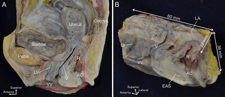
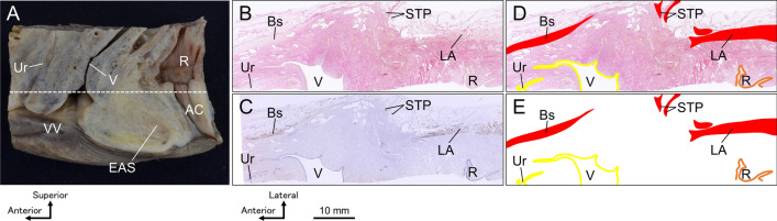
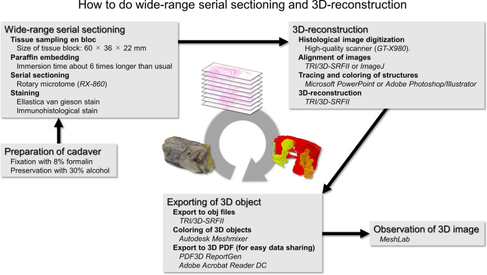
The present report presents details of the method for combining wide-range serial sectioning and 3D reconstruction using an adult cadaver. For several decades, anatomists have utilized a variety of non-destructive three-dimensional (3D) visualization methods to complement gross anatomical analysis methods. These include vascular casting for the visualization of vascular morphology and micro-CT for the visualization of bone morphology. However, these conventional methods are restricted by the properties and sizes of the target structures. Here, we introduce a method to conduct 3D reconstruction based on wide-range serial histological sections from adult cadavers, which overcomes previous restrictions. An attempt at 3D visualization of the female pelvic floor muscles provides a detailed description of the procedure. Supplemental video and 3D PDF files allow multifaceted observation of 3D images. Wide-range serial sectioning visualizes morphology beyond the scope of conventional methods, while 3D reconstruction enables non-destructive 3D visualization of any structure that can be observed on a histological section, including skeletal muscle, smooth muscle, ligaments, cartilage, connective tissue, blood vessels, nerves, lymph nodes, and glands. The novel combination of both methods is instrumental in meso-anatomy, a discipline intermediate between macro-anatomy and micro-anatomy.
期刊介绍:
The official English journal of the Japanese Association of Anatomists, Anatomical Science International (formerly titled Kaibogaku Zasshi) publishes original research articles dealing with morphological sciences.
Coverage in the journal includes molecular, cellular, histological and gross anatomical studies on humans and on normal and experimental animals, as well as functional morphological, biochemical, physiological and behavioral studies if they include morphological analysis.

 求助内容:
求助内容: 应助结果提醒方式:


