Mayyadah H Alabdely, Abdullah S Alolayan, Reem S Almaghrabi, Hail M Al-Abdely
{"title":"沙特阿拉伯某三级保健中心的脑褐丝酵母菌病。","authors":"Mayyadah H Alabdely, Abdullah S Alolayan, Reem S Almaghrabi, Hail M Al-Abdely","doi":"10.17712/nsj.2023.2.20220118","DOIUrl":null,"url":null,"abstract":"<p><strong>Objectives: </strong>To report cases of cerebral phaeohyphomycosis at a tertiary hospital in Riyadh, Saudi Arabia. Phaeohyphomycetes are a widely distributed group of fungi whose cell walls contain 1,8 dihydroxynaphthalene-melanin. Cerebral infections caused by these fungi are uncommon and primarily associated with neurotrophic phaeohyphomycetes.</p><p><strong>Methods: </strong>In January of 2020 we looked back to identify cases of culture-positive cerebral phaeohyphomycosis from our medical records at King Faisal Specialist Hospital and Research Center in Riyadh, Saudi Arabia. Data on demographics, potential risk factors, clinical presentation, treatment, and outcomes were analyzed.</p><p><strong>Results: </strong>Twelve cases of cerebral phaeohyphomycosis were identified, of which 4 were caused by Rhinocladiella mackenziei and the other 8 were caused by various phaeohyphomycetes. There were 2 cases caused by <i>Neoscytalidium dimidiatum</i>, and one case each caused by the following: <i>Acrophialophora fusispora</i>, <i>Chaetomium atrobrunneum</i>, <i>Exophiala dermatitidis</i>, <i>Exerohilum rostratum</i>, <i>Fonsecaea pedrosoi</i>, and <i>Cladophialophora bantiana</i>. Most patients (10 of 12) had underlying immunosuppression. <i>R. mackenziei</i> caused a brain-only infection manifesting as abscess formation. Four patients survived for more than a year after therapy. Surgical evacuation and triazole therapy with posaconazole or itraconazole, alone or in combination with other antifungal agents, were associated with success.</p><p><strong>Conclusion: </strong>Cerebral phaeohyphomycosis is an uncommon fungal infection that primarily affects immunocompromised patients and is associated with poor prognosis. <i>R. mackenziei</i> is the most prevalent fungus in our facility and has been linked to a universal mortality.</p>","PeriodicalId":19284,"journal":{"name":"Neurosciences","volume":null,"pages":null},"PeriodicalIF":1.2000,"publicationDate":"2023-04-01","publicationTypes":"Journal Article","fieldsOfStudy":null,"isOpenAccess":false,"openAccessPdf":"https://ftp.ncbi.nlm.nih.gov/pub/pmc/oa_pdf/98/db/Neurosciences-28-2-136.PMC10155477.pdf","citationCount":"0","resultStr":"{\"title\":\"Cerebral phaeohyphomycosis at a tertiary healthcare center in Saudi Arabia.\",\"authors\":\"Mayyadah H Alabdely, Abdullah S Alolayan, Reem S Almaghrabi, Hail M Al-Abdely\",\"doi\":\"10.17712/nsj.2023.2.20220118\",\"DOIUrl\":null,\"url\":null,\"abstract\":\"<p><strong>Objectives: </strong>To report cases of cerebral phaeohyphomycosis at a tertiary hospital in Riyadh, Saudi Arabia. Phaeohyphomycetes are a widely distributed group of fungi whose cell walls contain 1,8 dihydroxynaphthalene-melanin. Cerebral infections caused by these fungi are uncommon and primarily associated with neurotrophic phaeohyphomycetes.</p><p><strong>Methods: </strong>In January of 2020 we looked back to identify cases of culture-positive cerebral phaeohyphomycosis from our medical records at King Faisal Specialist Hospital and Research Center in Riyadh, Saudi Arabia. Data on demographics, potential risk factors, clinical presentation, treatment, and outcomes were analyzed.</p><p><strong>Results: </strong>Twelve cases of cerebral phaeohyphomycosis were identified, of which 4 were caused by Rhinocladiella mackenziei and the other 8 were caused by various phaeohyphomycetes. There were 2 cases caused by <i>Neoscytalidium dimidiatum</i>, and one case each caused by the following: <i>Acrophialophora fusispora</i>, <i>Chaetomium atrobrunneum</i>, <i>Exophiala dermatitidis</i>, <i>Exerohilum rostratum</i>, <i>Fonsecaea pedrosoi</i>, and <i>Cladophialophora bantiana</i>. Most patients (10 of 12) had underlying immunosuppression. <i>R. mackenziei</i> caused a brain-only infection manifesting as abscess formation. Four patients survived for more than a year after therapy. Surgical evacuation and triazole therapy with posaconazole or itraconazole, alone or in combination with other antifungal agents, were associated with success.</p><p><strong>Conclusion: </strong>Cerebral phaeohyphomycosis is an uncommon fungal infection that primarily affects immunocompromised patients and is associated with poor prognosis. <i>R. mackenziei</i> is the most prevalent fungus in our facility and has been linked to a universal mortality.</p>\",\"PeriodicalId\":19284,\"journal\":{\"name\":\"Neurosciences\",\"volume\":null,\"pages\":null},\"PeriodicalIF\":1.2000,\"publicationDate\":\"2023-04-01\",\"publicationTypes\":\"Journal Article\",\"fieldsOfStudy\":null,\"isOpenAccess\":false,\"openAccessPdf\":\"https://ftp.ncbi.nlm.nih.gov/pub/pmc/oa_pdf/98/db/Neurosciences-28-2-136.PMC10155477.pdf\",\"citationCount\":\"0\",\"resultStr\":null,\"platform\":\"Semanticscholar\",\"paperid\":null,\"PeriodicalName\":\"Neurosciences\",\"FirstCategoryId\":\"3\",\"ListUrlMain\":\"https://doi.org/10.17712/nsj.2023.2.20220118\",\"RegionNum\":4,\"RegionCategory\":\"医学\",\"ArticlePicture\":[],\"TitleCN\":null,\"AbstractTextCN\":null,\"PMCID\":null,\"EPubDate\":\"\",\"PubModel\":\"\",\"JCR\":\"Q4\",\"JCRName\":\"CLINICAL NEUROLOGY\",\"Score\":null,\"Total\":0}","platform":"Semanticscholar","paperid":null,"PeriodicalName":"Neurosciences","FirstCategoryId":"3","ListUrlMain":"https://doi.org/10.17712/nsj.2023.2.20220118","RegionNum":4,"RegionCategory":"医学","ArticlePicture":[],"TitleCN":null,"AbstractTextCN":null,"PMCID":null,"EPubDate":"","PubModel":"","JCR":"Q4","JCRName":"CLINICAL NEUROLOGY","Score":null,"Total":0}
Cerebral phaeohyphomycosis at a tertiary healthcare center in Saudi Arabia.
Objectives: To report cases of cerebral phaeohyphomycosis at a tertiary hospital in Riyadh, Saudi Arabia. Phaeohyphomycetes are a widely distributed group of fungi whose cell walls contain 1,8 dihydroxynaphthalene-melanin. Cerebral infections caused by these fungi are uncommon and primarily associated with neurotrophic phaeohyphomycetes.
Methods: In January of 2020 we looked back to identify cases of culture-positive cerebral phaeohyphomycosis from our medical records at King Faisal Specialist Hospital and Research Center in Riyadh, Saudi Arabia. Data on demographics, potential risk factors, clinical presentation, treatment, and outcomes were analyzed.
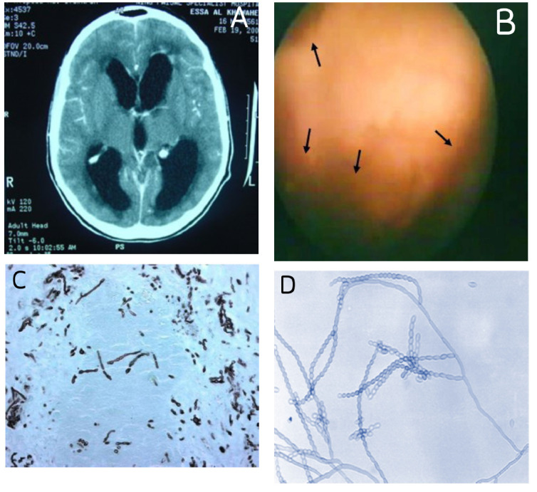
Results: Twelve cases of cerebral phaeohyphomycosis were identified, of which 4 were caused by Rhinocladiella mackenziei and the other 8 were caused by various phaeohyphomycetes. There were 2 cases caused by Neoscytalidium dimidiatum, and one case each caused by the following: Acrophialophora fusispora, Chaetomium atrobrunneum, Exophiala dermatitidis, Exerohilum rostratum, Fonsecaea pedrosoi, and Cladophialophora bantiana. Most patients (10 of 12) had underlying immunosuppression. R. mackenziei caused a brain-only infection manifesting as abscess formation. Four patients survived for more than a year after therapy. Surgical evacuation and triazole therapy with posaconazole or itraconazole, alone or in combination with other antifungal agents, were associated with success.
Conclusion: Cerebral phaeohyphomycosis is an uncommon fungal infection that primarily affects immunocompromised patients and is associated with poor prognosis. R. mackenziei is the most prevalent fungus in our facility and has been linked to a universal mortality.
期刊介绍:
Neurosciences is an open access, peer-reviewed, quarterly publication. Authors are invited to submit for publication articles reporting original work related to the nervous system, e.g., neurology, neurophysiology, neuroradiology, neurosurgery, neurorehabilitation, neurooncology, neuropsychiatry, and neurogenetics, etc. Basic research withclear clinical implications will also be considered. Review articles of current interest and high standard are welcomed for consideration. Prospective workshould not be backdated. There are also sections for Case Reports, Brief Communication, Correspondence, and medical news items. To promote continuous education, training, and learning, we include Clinical Images and MCQ’s. Highlights of international and regional meetings of interest, and specialized supplements will also be considered. All submissions must conform to the Uniform Requirements.
 求助内容:
求助内容: 应助结果提醒方式:


