Hiroaki Fukuzawa, Naoki Minoda, Mitsumasa Okamoto, Yudai Tsuruno, Aya Watanabe
{"title":"儿童阑尾、睾丸或附睾扭转的超声造影表现:一系列病例。","authors":"Hiroaki Fukuzawa, Naoki Minoda, Mitsumasa Okamoto, Yudai Tsuruno, Aya Watanabe","doi":"10.1007/s10396-023-01377-w","DOIUrl":null,"url":null,"abstract":"<p><strong>Purpose: </strong>Torsion of the appendix testis or epididymis is a cause of acute scrotum in children. Ultrasonography with color Doppler is the first-choice modality for diagnosis. However, this method requires skill and experience to make a diagnosis with confidence. Recently, contrast-enhanced ultrasonography for diagnosis in various fields has been reported. However, to our knowledge, there has been no report of this method being used to diagnose torsion of the appendix testis or epididymis. The purpose of this study was to retrospectively examine contrast-enhanced ultrasonographic findings in torsion of the appendix testis or epididymis.</p><p><strong>Methods: </strong>Patients who underwent contrast-enhanced ultrasonography for torsion of the appendix testis or epididymis at our institution between April 2010 and April 2023 were enrolled in this study (n = 12). Contrast-enhanced ultrasonography findings of the affected appendage and the testis parenchyma were examined retrospectively.</p><p><strong>Results: </strong>The parenchyma of the testes was notably enhanced in all the cases. However, 9 of the 12 cases showed that the appendage with torsion was not enhanced at all. In the remaining three cases, only slight enhancement was seen. Nevertheless, it was notably less than that of the parenchyma of the testis.</p><p><strong>Conclusion: </strong>Our findings indicated that contrast-enhanced ultrasonography may be an easy and reliable method for diagnosing torsion of the appendix testis or epididymis.</p>","PeriodicalId":50130,"journal":{"name":"Journal of Medical Ultrasonics","volume":null,"pages":null},"PeriodicalIF":1.9000,"publicationDate":"2024-01-01","publicationTypes":"Journal Article","fieldsOfStudy":null,"isOpenAccess":false,"openAccessPdf":"","citationCount":"0","resultStr":"{\"title\":\"Contrast-enhanced ultrasonographic findings in torsion of the appendix testis or epididymis in children: a case series.\",\"authors\":\"Hiroaki Fukuzawa, Naoki Minoda, Mitsumasa Okamoto, Yudai Tsuruno, Aya Watanabe\",\"doi\":\"10.1007/s10396-023-01377-w\",\"DOIUrl\":null,\"url\":null,\"abstract\":\"<p><strong>Purpose: </strong>Torsion of the appendix testis or epididymis is a cause of acute scrotum in children. Ultrasonography with color Doppler is the first-choice modality for diagnosis. However, this method requires skill and experience to make a diagnosis with confidence. Recently, contrast-enhanced ultrasonography for diagnosis in various fields has been reported. However, to our knowledge, there has been no report of this method being used to diagnose torsion of the appendix testis or epididymis. The purpose of this study was to retrospectively examine contrast-enhanced ultrasonographic findings in torsion of the appendix testis or epididymis.</p><p><strong>Methods: </strong>Patients who underwent contrast-enhanced ultrasonography for torsion of the appendix testis or epididymis at our institution between April 2010 and April 2023 were enrolled in this study (n = 12). Contrast-enhanced ultrasonography findings of the affected appendage and the testis parenchyma were examined retrospectively.</p><p><strong>Results: </strong>The parenchyma of the testes was notably enhanced in all the cases. However, 9 of the 12 cases showed that the appendage with torsion was not enhanced at all. In the remaining three cases, only slight enhancement was seen. Nevertheless, it was notably less than that of the parenchyma of the testis.</p><p><strong>Conclusion: </strong>Our findings indicated that contrast-enhanced ultrasonography may be an easy and reliable method for diagnosing torsion of the appendix testis or epididymis.</p>\",\"PeriodicalId\":50130,\"journal\":{\"name\":\"Journal of Medical Ultrasonics\",\"volume\":null,\"pages\":null},\"PeriodicalIF\":1.9000,\"publicationDate\":\"2024-01-01\",\"publicationTypes\":\"Journal Article\",\"fieldsOfStudy\":null,\"isOpenAccess\":false,\"openAccessPdf\":\"\",\"citationCount\":\"0\",\"resultStr\":null,\"platform\":\"Semanticscholar\",\"paperid\":null,\"PeriodicalName\":\"Journal of Medical Ultrasonics\",\"FirstCategoryId\":\"3\",\"ListUrlMain\":\"https://doi.org/10.1007/s10396-023-01377-w\",\"RegionNum\":4,\"RegionCategory\":\"医学\",\"ArticlePicture\":[],\"TitleCN\":null,\"AbstractTextCN\":null,\"PMCID\":null,\"EPubDate\":\"2023/10/21 0:00:00\",\"PubModel\":\"Epub\",\"JCR\":\"Q3\",\"JCRName\":\"RADIOLOGY, NUCLEAR MEDICINE & MEDICAL IMAGING\",\"Score\":null,\"Total\":0}","platform":"Semanticscholar","paperid":null,"PeriodicalName":"Journal of Medical Ultrasonics","FirstCategoryId":"3","ListUrlMain":"https://doi.org/10.1007/s10396-023-01377-w","RegionNum":4,"RegionCategory":"医学","ArticlePicture":[],"TitleCN":null,"AbstractTextCN":null,"PMCID":null,"EPubDate":"2023/10/21 0:00:00","PubModel":"Epub","JCR":"Q3","JCRName":"RADIOLOGY, NUCLEAR MEDICINE & MEDICAL IMAGING","Score":null,"Total":0}
Contrast-enhanced ultrasonographic findings in torsion of the appendix testis or epididymis in children: a case series.
Purpose: Torsion of the appendix testis or epididymis is a cause of acute scrotum in children. Ultrasonography with color Doppler is the first-choice modality for diagnosis. However, this method requires skill and experience to make a diagnosis with confidence. Recently, contrast-enhanced ultrasonography for diagnosis in various fields has been reported. However, to our knowledge, there has been no report of this method being used to diagnose torsion of the appendix testis or epididymis. The purpose of this study was to retrospectively examine contrast-enhanced ultrasonographic findings in torsion of the appendix testis or epididymis.
Methods: Patients who underwent contrast-enhanced ultrasonography for torsion of the appendix testis or epididymis at our institution between April 2010 and April 2023 were enrolled in this study (n = 12). Contrast-enhanced ultrasonography findings of the affected appendage and the testis parenchyma were examined retrospectively.
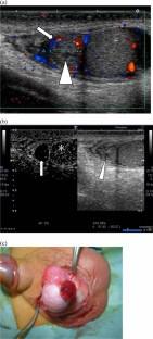
Results: The parenchyma of the testes was notably enhanced in all the cases. However, 9 of the 12 cases showed that the appendage with torsion was not enhanced at all. In the remaining three cases, only slight enhancement was seen. Nevertheless, it was notably less than that of the parenchyma of the testis.
Conclusion: Our findings indicated that contrast-enhanced ultrasonography may be an easy and reliable method for diagnosing torsion of the appendix testis or epididymis.
期刊介绍:
The Journal of Medical Ultrasonics is the official journal of the Japan Society of Ultrasonics in Medicine. The main purpose of the journal is to provide forum for the publication of papers documenting recent advances and new developments in the entire field of ultrasound in medicine and biology, encompassing both the medical and the engineering aspects of the science.The journal welcomes original articles, review articles, images, and letters to the editor.The journal also provides state-of-the-art information such as announcements from the boards and the committees of the society.
 求助内容:
求助内容: 应助结果提醒方式:


