{"title":"急性踝关节损伤:扭伤严重程度和辅助检查结果之间的关系。","authors":"Frederico Celestino Miranda, Eduardo Noda Kihara Filho, Marcelo Pires Prado, Laercio Alberto Rosemberg, Durval do Carmo Barros Santos, Atul Kumar Taneja","doi":"10.31744/einstein_journal/2023AO0162","DOIUrl":null,"url":null,"abstract":"<p><p>Miranda et al. reported a correlation between the significance of injuries to osseous, chondral, tendon, and ligamentous tissues in participants with low-grade versus high-grade acute ankle sprains. They demonstrated that participants with high-grade ankle sprains presented with shorter calcaneonavicular distances and increased rates of structural abnormalities compared to those with low-grade sprains. Special attention should be paid to acute ankle sprains in emergency settings to avoid failure in detecting severe injuries that could lead to chronic pain, impairment, or instability. Participants presenting acute ankle sprains (<15 days) were divided into low-grade versus high-grade sprain,according to the presence of a complete tear in at least one component of lateral ligament complex. High-grade ankle sprains group presented increased rates of medial malleolus bone bruise, deltoid ligament tears,extensor retinaculum lesions, and articular effusion. The calcaneonavicular distance was statistically shorter in patients with high-grade sprains (median, 3.0mm) when compared to those with low-grade sprains (median, 4.0mm) Objective: To correlate the significance of osseous, chondral, tendon, and ligamentous injuries with anatomical variations in low-grade versus high-grade acute ankle sprains.</p><p><strong>Methods: </strong>We retrospectively identified the magnetic resonance imaging findings of acute ankle sprains (<15 days). Participants with a history of previous sprains, arthritis, tumors, infections, or inflammatory conditions were excluded. Images were independently evaluated by two musculoskeletal radiologists and assessed for osseous, chondral, tendon, and ligamentous injuries and anatomical variations. Participants were divided into low-grade versus high-grade sprain groups, according to the presence of a complete tear in at least one component of the lateral ligament complex.</p><p><strong>Results: </strong>The final study group comprised 100 magnetic resonance images (mean age, 36 years), the majority of males (54%), the right ankle (52%), and a mean sprain duration of 5 days. Participants with high-grade sprains presented with increased rates of medial malleolus edema (p<0.001), moderate and large articular effusions (p=0.041), and shorter calcaneonavicular distance (p=0.008). Complete tears of the anterior talofibular ligament and calcaneofibular ligaments were observed in 100% and 51.2% of the participants in the High-Grade Group, respectively. The deltoid ligament complex was partially torn in this group (55.8% versus 8.8%, p<0.001). Extensor tendon retinaculum lesions occurred significantly more frequently in this group (41.9%) compared to the overall study population (23%) (p<0.001).</p><p><strong>Conclusion: </strong>Participants with high-grade ankle sprains presented with shorter calcaneonavicular distances and increased rates of medial malleolus edema, deltoid complex partial tears, extensor retinaculum lesions, and articular effusion.</p>","PeriodicalId":47359,"journal":{"name":"Einstein-Sao Paulo","volume":null,"pages":null},"PeriodicalIF":1.1000,"publicationDate":"2023-10-09","publicationTypes":"Journal Article","fieldsOfStudy":null,"isOpenAccess":false,"openAccessPdf":"https://www.ncbi.nlm.nih.gov/pmc/articles/PMC10519667/pdf/","citationCount":"0","resultStr":"{\"title\":\"Acute ankle injuries: association between sprain severity and ancillary findings.\",\"authors\":\"Frederico Celestino Miranda, Eduardo Noda Kihara Filho, Marcelo Pires Prado, Laercio Alberto Rosemberg, Durval do Carmo Barros Santos, Atul Kumar Taneja\",\"doi\":\"10.31744/einstein_journal/2023AO0162\",\"DOIUrl\":null,\"url\":null,\"abstract\":\"<p><p>Miranda et al. reported a correlation between the significance of injuries to osseous, chondral, tendon, and ligamentous tissues in participants with low-grade versus high-grade acute ankle sprains. They demonstrated that participants with high-grade ankle sprains presented with shorter calcaneonavicular distances and increased rates of structural abnormalities compared to those with low-grade sprains. Special attention should be paid to acute ankle sprains in emergency settings to avoid failure in detecting severe injuries that could lead to chronic pain, impairment, or instability. Participants presenting acute ankle sprains (<15 days) were divided into low-grade versus high-grade sprain,according to the presence of a complete tear in at least one component of lateral ligament complex. High-grade ankle sprains group presented increased rates of medial malleolus bone bruise, deltoid ligament tears,extensor retinaculum lesions, and articular effusion. The calcaneonavicular distance was statistically shorter in patients with high-grade sprains (median, 3.0mm) when compared to those with low-grade sprains (median, 4.0mm) Objective: To correlate the significance of osseous, chondral, tendon, and ligamentous injuries with anatomical variations in low-grade versus high-grade acute ankle sprains.</p><p><strong>Methods: </strong>We retrospectively identified the magnetic resonance imaging findings of acute ankle sprains (<15 days). Participants with a history of previous sprains, arthritis, tumors, infections, or inflammatory conditions were excluded. Images were independently evaluated by two musculoskeletal radiologists and assessed for osseous, chondral, tendon, and ligamentous injuries and anatomical variations. Participants were divided into low-grade versus high-grade sprain groups, according to the presence of a complete tear in at least one component of the lateral ligament complex.</p><p><strong>Results: </strong>The final study group comprised 100 magnetic resonance images (mean age, 36 years), the majority of males (54%), the right ankle (52%), and a mean sprain duration of 5 days. Participants with high-grade sprains presented with increased rates of medial malleolus edema (p<0.001), moderate and large articular effusions (p=0.041), and shorter calcaneonavicular distance (p=0.008). Complete tears of the anterior talofibular ligament and calcaneofibular ligaments were observed in 100% and 51.2% of the participants in the High-Grade Group, respectively. The deltoid ligament complex was partially torn in this group (55.8% versus 8.8%, p<0.001). Extensor tendon retinaculum lesions occurred significantly more frequently in this group (41.9%) compared to the overall study population (23%) (p<0.001).</p><p><strong>Conclusion: </strong>Participants with high-grade ankle sprains presented with shorter calcaneonavicular distances and increased rates of medial malleolus edema, deltoid complex partial tears, extensor retinaculum lesions, and articular effusion.</p>\",\"PeriodicalId\":47359,\"journal\":{\"name\":\"Einstein-Sao Paulo\",\"volume\":null,\"pages\":null},\"PeriodicalIF\":1.1000,\"publicationDate\":\"2023-10-09\",\"publicationTypes\":\"Journal Article\",\"fieldsOfStudy\":null,\"isOpenAccess\":false,\"openAccessPdf\":\"https://www.ncbi.nlm.nih.gov/pmc/articles/PMC10519667/pdf/\",\"citationCount\":\"0\",\"resultStr\":null,\"platform\":\"Semanticscholar\",\"paperid\":null,\"PeriodicalName\":\"Einstein-Sao Paulo\",\"FirstCategoryId\":\"1085\",\"ListUrlMain\":\"https://doi.org/10.31744/einstein_journal/2023AO0162\",\"RegionNum\":0,\"RegionCategory\":null,\"ArticlePicture\":[],\"TitleCN\":null,\"AbstractTextCN\":null,\"PMCID\":null,\"EPubDate\":\"2023/1/1 0:00:00\",\"PubModel\":\"eCollection\",\"JCR\":\"Q2\",\"JCRName\":\"MEDICINE, GENERAL & INTERNAL\",\"Score\":null,\"Total\":0}","platform":"Semanticscholar","paperid":null,"PeriodicalName":"Einstein-Sao Paulo","FirstCategoryId":"1085","ListUrlMain":"https://doi.org/10.31744/einstein_journal/2023AO0162","RegionNum":0,"RegionCategory":null,"ArticlePicture":[],"TitleCN":null,"AbstractTextCN":null,"PMCID":null,"EPubDate":"2023/1/1 0:00:00","PubModel":"eCollection","JCR":"Q2","JCRName":"MEDICINE, GENERAL & INTERNAL","Score":null,"Total":0}
Acute ankle injuries: association between sprain severity and ancillary findings.
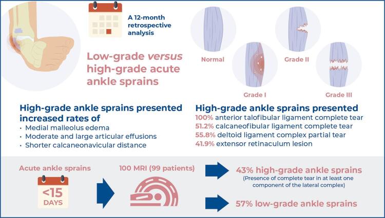
Miranda et al. reported a correlation between the significance of injuries to osseous, chondral, tendon, and ligamentous tissues in participants with low-grade versus high-grade acute ankle sprains. They demonstrated that participants with high-grade ankle sprains presented with shorter calcaneonavicular distances and increased rates of structural abnormalities compared to those with low-grade sprains. Special attention should be paid to acute ankle sprains in emergency settings to avoid failure in detecting severe injuries that could lead to chronic pain, impairment, or instability. Participants presenting acute ankle sprains (<15 days) were divided into low-grade versus high-grade sprain,according to the presence of a complete tear in at least one component of lateral ligament complex. High-grade ankle sprains group presented increased rates of medial malleolus bone bruise, deltoid ligament tears,extensor retinaculum lesions, and articular effusion. The calcaneonavicular distance was statistically shorter in patients with high-grade sprains (median, 3.0mm) when compared to those with low-grade sprains (median, 4.0mm) Objective: To correlate the significance of osseous, chondral, tendon, and ligamentous injuries with anatomical variations in low-grade versus high-grade acute ankle sprains.
Methods: We retrospectively identified the magnetic resonance imaging findings of acute ankle sprains (<15 days). Participants with a history of previous sprains, arthritis, tumors, infections, or inflammatory conditions were excluded. Images were independently evaluated by two musculoskeletal radiologists and assessed for osseous, chondral, tendon, and ligamentous injuries and anatomical variations. Participants were divided into low-grade versus high-grade sprain groups, according to the presence of a complete tear in at least one component of the lateral ligament complex.
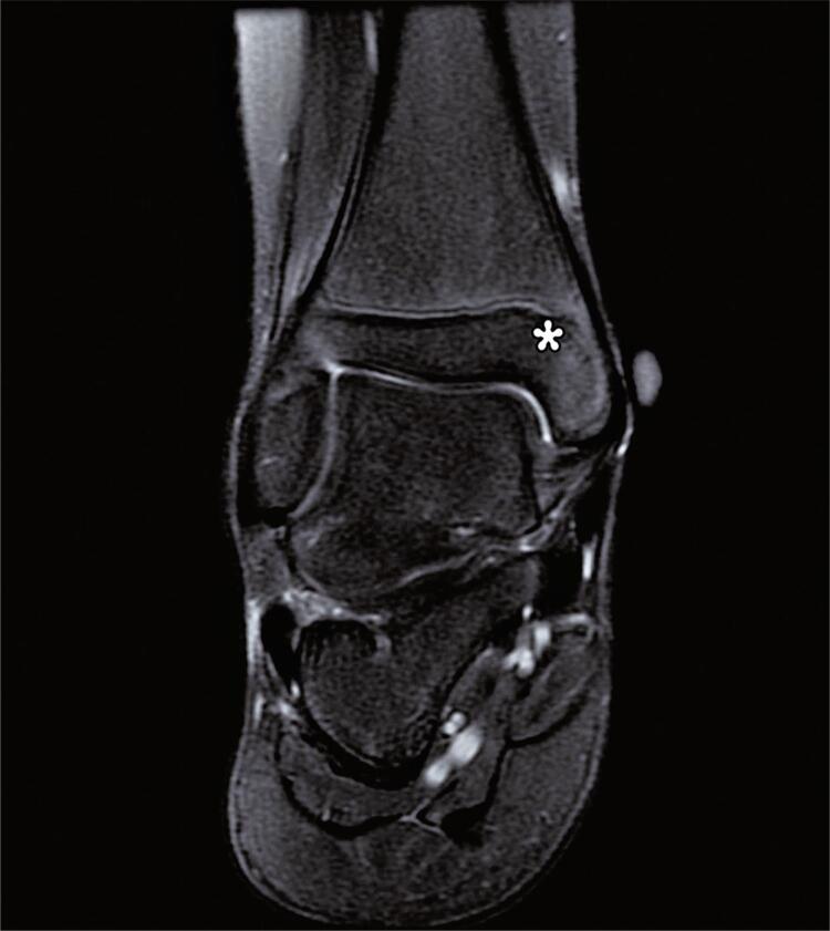
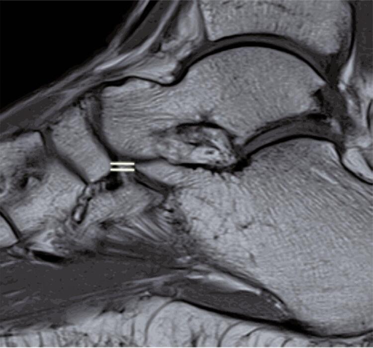
Results: The final study group comprised 100 magnetic resonance images (mean age, 36 years), the majority of males (54%), the right ankle (52%), and a mean sprain duration of 5 days. Participants with high-grade sprains presented with increased rates of medial malleolus edema (p<0.001), moderate and large articular effusions (p=0.041), and shorter calcaneonavicular distance (p=0.008). Complete tears of the anterior talofibular ligament and calcaneofibular ligaments were observed in 100% and 51.2% of the participants in the High-Grade Group, respectively. The deltoid ligament complex was partially torn in this group (55.8% versus 8.8%, p<0.001). Extensor tendon retinaculum lesions occurred significantly more frequently in this group (41.9%) compared to the overall study population (23%) (p<0.001).
Conclusion: Participants with high-grade ankle sprains presented with shorter calcaneonavicular distances and increased rates of medial malleolus edema, deltoid complex partial tears, extensor retinaculum lesions, and articular effusion.

 求助内容:
求助内容: 应助结果提醒方式:


