Omar Al-Louzi, Vijay Letchuman, Sargis Manukyan, Erin S Beck, Snehashis Roy, Joan Ohayon, Dzung L Pham, Irene Cortese, Pascal Sati, Daniel S Reich
{"title":"多发性硬化症新发病灶的中心静脉征象:一项为期3年的纵向研究。","authors":"Omar Al-Louzi, Vijay Letchuman, Sargis Manukyan, Erin S Beck, Snehashis Roy, Joan Ohayon, Dzung L Pham, Irene Cortese, Pascal Sati, Daniel S Reich","doi":"10.1212/NXI.0000000000001120","DOIUrl":null,"url":null,"abstract":"<p><strong>Background and objectives: </strong>The central vein sign (CVS), a central linear hypointensity within lesions on T2*-weighted imaging, has been established as a sensitive and specific biomarker for the diagnosis of multiple sclerosis (MS). However, the CVS has not yet been comprehensively studied in newly developing MS lesions. We aimed to identify the CVS profiles of new white matter lesions in patients with MS followed over time and investigate demographic and clinical risk factors associated with new CVS+ or CVS- lesion development.</p><p><strong>Methods: </strong>In this retrospective longitudinal cohort study, adults from the NIH MS Natural History Study were considered for inclusion. Participants with new T2 or enhancing lesions were identified through review of the radiology report and/or longitudinal subtraction imaging. Each new lesion was evaluated for the CVS. Clinical characteristics were identified through chart review.</p><p><strong>Results: </strong>A total of 153 adults (95 relapsing-remitting MS, 27 secondary progressive MS, 16 primary progressive MS, 5 clinically isolated syndrome, and 10 healthy; 67% female) were included. Of this cohort, 96 had at least 1 new T2 or contrast-enhancing lesion during median 3.1 years (Q1-Q3: 0.7-6.3) of follow-up; lesions eligible for CVS evaluation were found in 62 (65%). Of 233 new CVS-eligible lesions, 159 (68%) were CVS+, with 30 (48%) individuals having only CVS+, 12 (19%) only CVS-, and 20 (32%) both CVS+ and CVS- lesions. In gadolinium-enhancing (Gd+) lesions, the CVS+ percentage increased from 102/152 (67%) at the first time point where the lesion was observed, to 92/114 (82%) after a median follow-up of 2.8 years. Younger age (OR = 0.5 per 10-year increase, 95% CI = 0.3-0.8) and higher CVS+ percentage at baseline (OR = 1.4 per 10% increase, 95% CI = 1.1-1.9) were associated with increased likelihood of new CVS+ lesion development.</p><p><strong>Discussion: </strong>In a cohort of adults with MS followed over a median duration of 3 years, most newly developing T2 or enhancing lesions were CVS+ (68%), and nearly half (48%) developed new CVS+ lesions only. Importantly, the effects of edema and T2 signal changes can obscure small veins in Gd+ lesions; therefore, caution and follow-up is necessary when determining their CVS status.</p><p><strong>Trial registration information: </strong>Clinical trial registration number NCT00001248.</p><p><strong>Classification of evidence: </strong>This study provides Class III evidence that younger age and higher CVS+ percentage at baseline are associated with new CVS+ lesion development.</p>","PeriodicalId":520720,"journal":{"name":"Neurology(R) neuroimmunology & neuroinflammation","volume":" ","pages":""},"PeriodicalIF":7.5000,"publicationDate":"2022-01-13","publicationTypes":"Journal Article","fieldsOfStudy":null,"isOpenAccess":false,"openAccessPdf":"https://ftp.ncbi.nlm.nih.gov/pub/pmc/oa_pdf/09/6d/NEURIMMINFL2021039347.PMC8759076.pdf","citationCount":"4","resultStr":"{\"title\":\"Central Vein Sign Profile of Newly Developing Lesions in Multiple Sclerosis: A 3-Year Longitudinal Study.\",\"authors\":\"Omar Al-Louzi, Vijay Letchuman, Sargis Manukyan, Erin S Beck, Snehashis Roy, Joan Ohayon, Dzung L Pham, Irene Cortese, Pascal Sati, Daniel S Reich\",\"doi\":\"10.1212/NXI.0000000000001120\",\"DOIUrl\":null,\"url\":null,\"abstract\":\"<p><strong>Background and objectives: </strong>The central vein sign (CVS), a central linear hypointensity within lesions on T2*-weighted imaging, has been established as a sensitive and specific biomarker for the diagnosis of multiple sclerosis (MS). However, the CVS has not yet been comprehensively studied in newly developing MS lesions. We aimed to identify the CVS profiles of new white matter lesions in patients with MS followed over time and investigate demographic and clinical risk factors associated with new CVS+ or CVS- lesion development.</p><p><strong>Methods: </strong>In this retrospective longitudinal cohort study, adults from the NIH MS Natural History Study were considered for inclusion. Participants with new T2 or enhancing lesions were identified through review of the radiology report and/or longitudinal subtraction imaging. Each new lesion was evaluated for the CVS. Clinical characteristics were identified through chart review.</p><p><strong>Results: </strong>A total of 153 adults (95 relapsing-remitting MS, 27 secondary progressive MS, 16 primary progressive MS, 5 clinically isolated syndrome, and 10 healthy; 67% female) were included. Of this cohort, 96 had at least 1 new T2 or contrast-enhancing lesion during median 3.1 years (Q1-Q3: 0.7-6.3) of follow-up; lesions eligible for CVS evaluation were found in 62 (65%). Of 233 new CVS-eligible lesions, 159 (68%) were CVS+, with 30 (48%) individuals having only CVS+, 12 (19%) only CVS-, and 20 (32%) both CVS+ and CVS- lesions. In gadolinium-enhancing (Gd+) lesions, the CVS+ percentage increased from 102/152 (67%) at the first time point where the lesion was observed, to 92/114 (82%) after a median follow-up of 2.8 years. Younger age (OR = 0.5 per 10-year increase, 95% CI = 0.3-0.8) and higher CVS+ percentage at baseline (OR = 1.4 per 10% increase, 95% CI = 1.1-1.9) were associated with increased likelihood of new CVS+ lesion development.</p><p><strong>Discussion: </strong>In a cohort of adults with MS followed over a median duration of 3 years, most newly developing T2 or enhancing lesions were CVS+ (68%), and nearly half (48%) developed new CVS+ lesions only. Importantly, the effects of edema and T2 signal changes can obscure small veins in Gd+ lesions; therefore, caution and follow-up is necessary when determining their CVS status.</p><p><strong>Trial registration information: </strong>Clinical trial registration number NCT00001248.</p><p><strong>Classification of evidence: </strong>This study provides Class III evidence that younger age and higher CVS+ percentage at baseline are associated with new CVS+ lesion development.</p>\",\"PeriodicalId\":520720,\"journal\":{\"name\":\"Neurology(R) neuroimmunology & neuroinflammation\",\"volume\":\" \",\"pages\":\"\"},\"PeriodicalIF\":7.5000,\"publicationDate\":\"2022-01-13\",\"publicationTypes\":\"Journal Article\",\"fieldsOfStudy\":null,\"isOpenAccess\":false,\"openAccessPdf\":\"https://ftp.ncbi.nlm.nih.gov/pub/pmc/oa_pdf/09/6d/NEURIMMINFL2021039347.PMC8759076.pdf\",\"citationCount\":\"4\",\"resultStr\":null,\"platform\":\"Semanticscholar\",\"paperid\":null,\"PeriodicalName\":\"Neurology(R) neuroimmunology & neuroinflammation\",\"FirstCategoryId\":\"3\",\"ListUrlMain\":\"https://doi.org/10.1212/NXI.0000000000001120\",\"RegionNum\":0,\"RegionCategory\":null,\"ArticlePicture\":[],\"TitleCN\":null,\"AbstractTextCN\":null,\"PMCID\":null,\"EPubDate\":\"2022/3/1 0:00:00\",\"PubModel\":\"Print\",\"JCR\":\"\",\"JCRName\":\"\",\"Score\":null,\"Total\":0}","platform":"Semanticscholar","paperid":null,"PeriodicalName":"Neurology(R) neuroimmunology & neuroinflammation","FirstCategoryId":"3","ListUrlMain":"https://doi.org/10.1212/NXI.0000000000001120","RegionNum":0,"RegionCategory":null,"ArticlePicture":[],"TitleCN":null,"AbstractTextCN":null,"PMCID":null,"EPubDate":"2022/3/1 0:00:00","PubModel":"Print","JCR":"","JCRName":"","Score":null,"Total":0}
Central Vein Sign Profile of Newly Developing Lesions in Multiple Sclerosis: A 3-Year Longitudinal Study.
Background and objectives: The central vein sign (CVS), a central linear hypointensity within lesions on T2*-weighted imaging, has been established as a sensitive and specific biomarker for the diagnosis of multiple sclerosis (MS). However, the CVS has not yet been comprehensively studied in newly developing MS lesions. We aimed to identify the CVS profiles of new white matter lesions in patients with MS followed over time and investigate demographic and clinical risk factors associated with new CVS+ or CVS- lesion development.
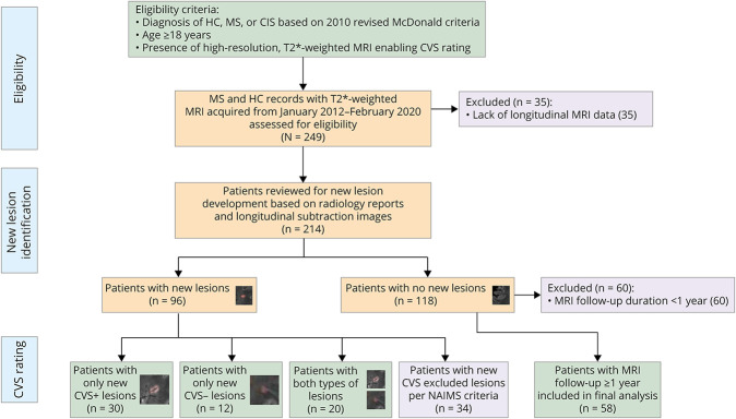
Methods: In this retrospective longitudinal cohort study, adults from the NIH MS Natural History Study were considered for inclusion. Participants with new T2 or enhancing lesions were identified through review of the radiology report and/or longitudinal subtraction imaging. Each new lesion was evaluated for the CVS. Clinical characteristics were identified through chart review.
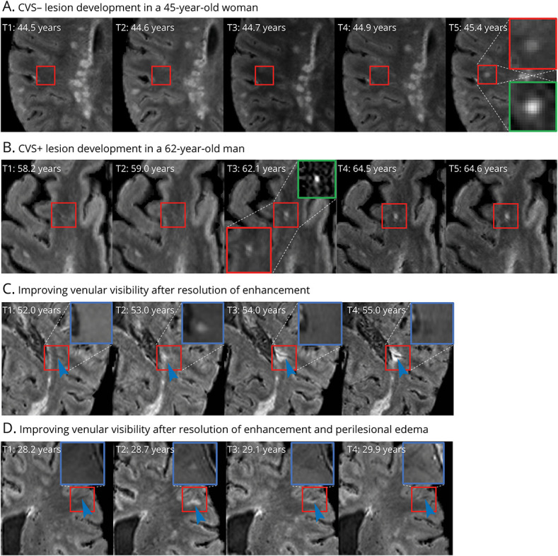
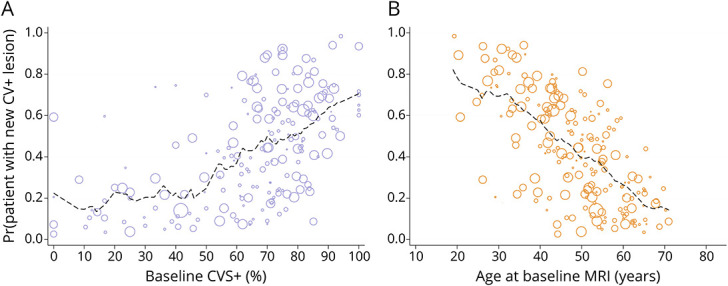
Results: A total of 153 adults (95 relapsing-remitting MS, 27 secondary progressive MS, 16 primary progressive MS, 5 clinically isolated syndrome, and 10 healthy; 67% female) were included. Of this cohort, 96 had at least 1 new T2 or contrast-enhancing lesion during median 3.1 years (Q1-Q3: 0.7-6.3) of follow-up; lesions eligible for CVS evaluation were found in 62 (65%). Of 233 new CVS-eligible lesions, 159 (68%) were CVS+, with 30 (48%) individuals having only CVS+, 12 (19%) only CVS-, and 20 (32%) both CVS+ and CVS- lesions. In gadolinium-enhancing (Gd+) lesions, the CVS+ percentage increased from 102/152 (67%) at the first time point where the lesion was observed, to 92/114 (82%) after a median follow-up of 2.8 years. Younger age (OR = 0.5 per 10-year increase, 95% CI = 0.3-0.8) and higher CVS+ percentage at baseline (OR = 1.4 per 10% increase, 95% CI = 1.1-1.9) were associated with increased likelihood of new CVS+ lesion development.
Discussion: In a cohort of adults with MS followed over a median duration of 3 years, most newly developing T2 or enhancing lesions were CVS+ (68%), and nearly half (48%) developed new CVS+ lesions only. Importantly, the effects of edema and T2 signal changes can obscure small veins in Gd+ lesions; therefore, caution and follow-up is necessary when determining their CVS status.
Trial registration information: Clinical trial registration number NCT00001248.
Classification of evidence: This study provides Class III evidence that younger age and higher CVS+ percentage at baseline are associated with new CVS+ lesion development.

 求助内容:
求助内容: 应助结果提醒方式:


