{"title":"核形态学在预测弥漫性浸润胶质瘤分级中的应用。","authors":"Dibyajyoti Boruah, Prabal Deb","doi":"10.1155/2013/760653","DOIUrl":null,"url":null,"abstract":"<p><p>Introduction. The ability to reliably differentiate neoplastic from nonneoplastic specimen and ascertain the tumour grade of diffusely infiltrating gliomas (DIGs) is often challenging. Aims and Objective. To evaluate utility of image morphometry in identifying DIG areas and to predict tumour grade. Materials and Methods. Image morphometry was used to analyze the following nuclear features of 30 DIGs and 10 controls (CG): major axis of nucleus (MAJX), minor axis of nucleus (MINX), nuclear area (NA), nuclear perimeter (NP), nuclear roundness (NR), nuclear density (ND), and percentage of total nuclear area (%TNA). Results. Statistically significant differences in all parameters, except NR, were observed between all groups, with strong positive correlation with tumour grade (r > 0.7). The mean values were maximum for HGG and minimum for CG. For NR, the difference between CG/HGG was statistically significant, unlike CG/LGG and LGG/HGG. It was observed that NA distributions for CG were nearly Gaussian type with smaller range, while gliomas displayed erratic pattern with larger range. NA and NP exhibited strong positive correlation with ND. Conclusion. Image morphometry has immense potential in being a powerful tool to distinguish normal from neoplastic tissue and also to differentiate LGG from HGG cases, especially in tiny stereotactic biopsies. </p>","PeriodicalId":89399,"journal":{"name":"ISRN oncology","volume":"2013 ","pages":"760653"},"PeriodicalIF":0.0000,"publicationDate":"2013-08-26","publicationTypes":"Journal Article","fieldsOfStudy":null,"isOpenAccess":false,"openAccessPdf":"https://sci-hub-pdf.com/10.1155/2013/760653","citationCount":"8","resultStr":"{\"title\":\"Utility of nuclear morphometry in predicting grades of diffusely infiltrating gliomas.\",\"authors\":\"Dibyajyoti Boruah, Prabal Deb\",\"doi\":\"10.1155/2013/760653\",\"DOIUrl\":null,\"url\":null,\"abstract\":\"<p><p>Introduction. The ability to reliably differentiate neoplastic from nonneoplastic specimen and ascertain the tumour grade of diffusely infiltrating gliomas (DIGs) is often challenging. Aims and Objective. To evaluate utility of image morphometry in identifying DIG areas and to predict tumour grade. Materials and Methods. Image morphometry was used to analyze the following nuclear features of 30 DIGs and 10 controls (CG): major axis of nucleus (MAJX), minor axis of nucleus (MINX), nuclear area (NA), nuclear perimeter (NP), nuclear roundness (NR), nuclear density (ND), and percentage of total nuclear area (%TNA). Results. Statistically significant differences in all parameters, except NR, were observed between all groups, with strong positive correlation with tumour grade (r > 0.7). The mean values were maximum for HGG and minimum for CG. For NR, the difference between CG/HGG was statistically significant, unlike CG/LGG and LGG/HGG. It was observed that NA distributions for CG were nearly Gaussian type with smaller range, while gliomas displayed erratic pattern with larger range. NA and NP exhibited strong positive correlation with ND. Conclusion. Image morphometry has immense potential in being a powerful tool to distinguish normal from neoplastic tissue and also to differentiate LGG from HGG cases, especially in tiny stereotactic biopsies. </p>\",\"PeriodicalId\":89399,\"journal\":{\"name\":\"ISRN oncology\",\"volume\":\"2013 \",\"pages\":\"760653\"},\"PeriodicalIF\":0.0000,\"publicationDate\":\"2013-08-26\",\"publicationTypes\":\"Journal Article\",\"fieldsOfStudy\":null,\"isOpenAccess\":false,\"openAccessPdf\":\"https://sci-hub-pdf.com/10.1155/2013/760653\",\"citationCount\":\"8\",\"resultStr\":null,\"platform\":\"Semanticscholar\",\"paperid\":null,\"PeriodicalName\":\"ISRN oncology\",\"FirstCategoryId\":\"1085\",\"ListUrlMain\":\"https://doi.org/10.1155/2013/760653\",\"RegionNum\":0,\"RegionCategory\":null,\"ArticlePicture\":[],\"TitleCN\":null,\"AbstractTextCN\":null,\"PMCID\":null,\"EPubDate\":\"2013/1/1 0:00:00\",\"PubModel\":\"eCollection\",\"JCR\":\"\",\"JCRName\":\"\",\"Score\":null,\"Total\":0}","platform":"Semanticscholar","paperid":null,"PeriodicalName":"ISRN oncology","FirstCategoryId":"1085","ListUrlMain":"https://doi.org/10.1155/2013/760653","RegionNum":0,"RegionCategory":null,"ArticlePicture":[],"TitleCN":null,"AbstractTextCN":null,"PMCID":null,"EPubDate":"2013/1/1 0:00:00","PubModel":"eCollection","JCR":"","JCRName":"","Score":null,"Total":0}
Utility of nuclear morphometry in predicting grades of diffusely infiltrating gliomas.
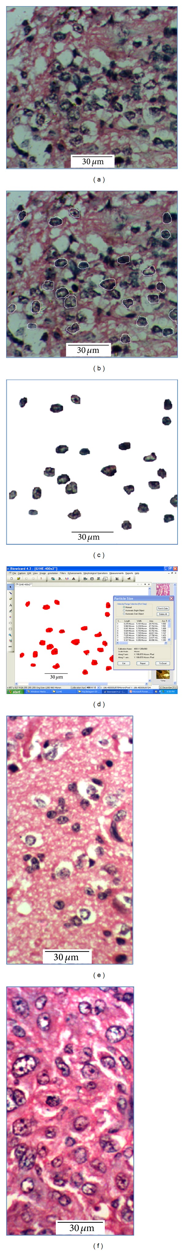
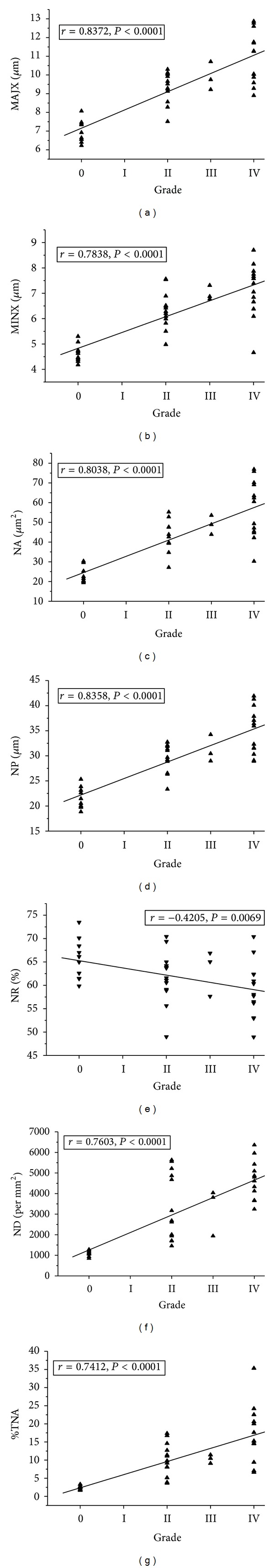
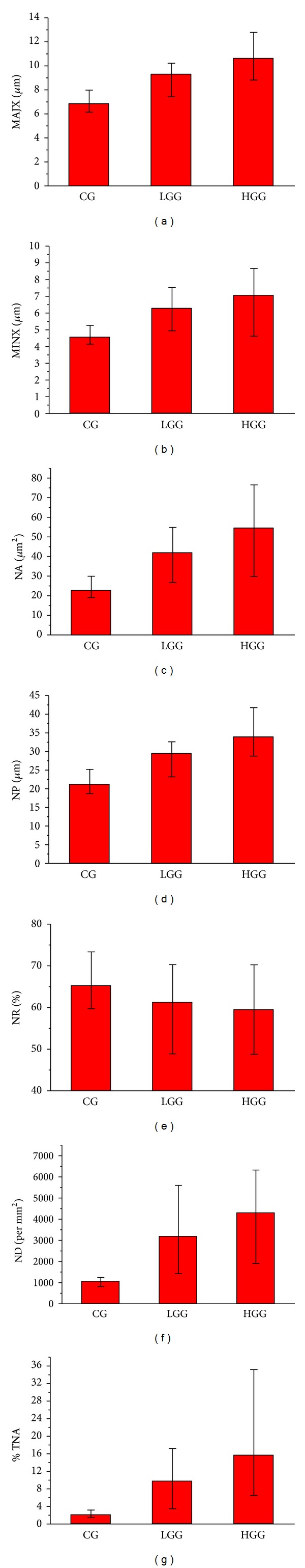
Introduction. The ability to reliably differentiate neoplastic from nonneoplastic specimen and ascertain the tumour grade of diffusely infiltrating gliomas (DIGs) is often challenging. Aims and Objective. To evaluate utility of image morphometry in identifying DIG areas and to predict tumour grade. Materials and Methods. Image morphometry was used to analyze the following nuclear features of 30 DIGs and 10 controls (CG): major axis of nucleus (MAJX), minor axis of nucleus (MINX), nuclear area (NA), nuclear perimeter (NP), nuclear roundness (NR), nuclear density (ND), and percentage of total nuclear area (%TNA). Results. Statistically significant differences in all parameters, except NR, were observed between all groups, with strong positive correlation with tumour grade (r > 0.7). The mean values were maximum for HGG and minimum for CG. For NR, the difference between CG/HGG was statistically significant, unlike CG/LGG and LGG/HGG. It was observed that NA distributions for CG were nearly Gaussian type with smaller range, while gliomas displayed erratic pattern with larger range. NA and NP exhibited strong positive correlation with ND. Conclusion. Image morphometry has immense potential in being a powerful tool to distinguish normal from neoplastic tissue and also to differentiate LGG from HGG cases, especially in tiny stereotactic biopsies.

 求助内容:
求助内容: 应助结果提醒方式:


